Repair of the tympanic membrane with urinary bladder matrix
- PMID: 19358244
- PMCID: PMC3003594
- DOI: 10.1002/lary.20233
Repair of the tympanic membrane with urinary bladder matrix
Abstract
Objectives/hypothesis: To test urinary bladder matrix (UBM) as a potential treatment for tympanic membrane (TM) healing and regeneration.
Study design: This prospective pilot study was designed to provide both qualitative and semiquantitative assessment of temporal and spatial healing events in the chinchilla model of chronic TM perforations with and without UBM patching.
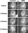
Methods: Bilateral myringotomies were performed and repeated as necessary to create subtotal perforations over an 8-week period. Myringoplasty was then performed, with left TMs serving as controls and right TMs receiving UBM patches. TMs were excised at 4 weeks, 8 weeks, and 12 weeks. Fixed tissue samples were characterized for gross morphology, then processed for microscopic evaluation.
Results: Chronic perforations were maintained with one or more repeated myringotomies. Although both control and patched TMs were thicker than native tissue, patched TMs were transparent and uniform in thickness without any inclusions. UBM patches were readily degraded and replaced by newly deposited and organized host tissue that recapitulated the native TM layers.
Conclusions: UBM scaffolds were an effective biological scaffold for TM closure and tissue remodeling, leading to thicker than normal anatomy but otherwise normal morphology. Future studies are required to determine functional and temporal outcomes as well as alternative patch orientations. The results show particular promise as a superior alternative means of reconstructing not only chronic TM perforations but also dimeric TMs associated with retraction pockets and atelectasis. Laryngoscope, 2009.
Figures


Similar articles
-
Poly(glycerol sebacate)-engineered plugs to repair chronic tympanic membrane perforations in a chinchilla model.Otolaryngol Head Neck Surg. 2010 Jul;143(1):127-33. doi: 10.1016/j.otohns.2010.01.025. Otolaryngol Head Neck Surg. 2010. PMID: 20620631
-
Tympanic membrane repair using silk fibroin and acellular collagen scaffolds.Laryngoscope. 2013 Aug;123(8):1976-82. doi: 10.1002/lary.23940. Epub 2013 Mar 27. Laryngoscope. 2013. PMID: 23536496
-
An experimental study on tympanic membrane reconstruction with acellular dermal matrix.Acta Otolaryngol. 2012 Dec;132(12):1266-70. doi: 10.3109/00016489.2012.701327. Epub 2012 Jul 25. Acta Otolaryngol. 2012. PMID: 22831646
-
Application of mesenchymal stem cell for tympanic membrane regeneration by tissue engineering approach.Int J Pediatr Otorhinolaryngol. 2020 Jun;133:109969. doi: 10.1016/j.ijporl.2020.109969. Epub 2020 Feb 25. Int J Pediatr Otorhinolaryngol. 2020. PMID: 32126416 Review.
-
A moist edge environment aids the regeneration of traumatic tympanic membrane perforations.J Laryngol Otol. 2017 Jul;131(7):564-571. doi: 10.1017/S0022215117001001. Epub 2017 May 15. J Laryngol Otol. 2017. PMID: 28502255 Review.
Cited by
-
Effects of topical applications of porcine acellular urinary bladder matrix and Centella asiatica extract on oral wound healing in a rat model.Clin Oral Investig. 2019 May;23(5):2083-2095. doi: 10.1007/s00784-018-2620-x. Epub 2018 Sep 24. Clin Oral Investig. 2019. PMID: 30251055
-
Scaffolds for tympanic membrane regeneration in rats.Tissue Eng Part A. 2013 Mar;19(5-6):657-68. doi: 10.1089/ten.TEA.2012.0053. Epub 2012 Dec 10. Tissue Eng Part A. 2013. PMID: 23092139 Free PMC article.
-
[Sugarcane biopolymer membrane: experimental evaluation in the middle ear].Braz J Otorhinolaryngol. 2011 Jan-Feb;77(1):44-50. doi: 10.1590/S1808-86942011000100008. Braz J Otorhinolaryngol. 2011. PMID: 21340188 Free PMC article.
-
The effect of detergents on the basement membrane complex of a biologic scaffold material.Acta Biomater. 2014 Jan;10(1):183-93. doi: 10.1016/j.actbio.2013.09.006. Epub 2013 Sep 18. Acta Biomater. 2014. PMID: 24055455 Free PMC article.
-
Animal models of chronic tympanic membrane perforation: in response to plasminogen initiates and potentiates the healing of acute and chronic tympanic membrane perforations in mice.Clin Transl Med. 2014 Mar 26;3(1):5. doi: 10.1186/2001-1326-3-5. Clin Transl Med. 2014. PMID: 24669846 Free PMC article.
References
-
- Ross MH, Reith EJ, Romrell LJ. A Text and Atlas. 2. Baltimore, MD: Williams & Wilkins; 1989. Histology.
-
- Johnson AP, Smallman LA, Kent SE. The mechanism of healing of tympanic membrane perforations. Acta Otolaryngol. 1990;109:406–415. - PubMed
-
- O’Reilly RC, Goldman SA, Widner SA, Cass SP. Creating a stable tympanic membrane perforation using mitomycin C. Otolaryngol Head Neck Surg. 2001;124:40–45. - PubMed
-
- Berger G. Nature of spontaneous tympanic membrane perforation in acute otitis media in children. J Laryngol Otol. 1989;103:1150–1153. - PubMed
-
- Lindeman P, Edstrom S, Granstrom G, et al. Acute traumatic tympanic membrane perforations. Arch Otolaryngol. 1987;113:1285–1287. - PubMed
Publication types
MeSH terms
Grants and funding
LinkOut - more resources
Full Text Sources
Other Literature Sources

