. 2009 May;46(5):728-38.
doi: 10.1016/j.yjmcc.2009.01.017.

Thymosin beta4 mediated PKC activation is essential to initiate the embryonic coronary developmental program and epicardial progenitor cell activation in adult mice in vivo

Affiliations

Thymosin beta4 mediated PKC activation is essential to initiate the embryonic coronary developmental program and epicardial progenitor cell activation in adult mice in vivo

Ildiko Bock-Marquette et al. J Mol Cell Cardiol. 2009 May.

Abstract

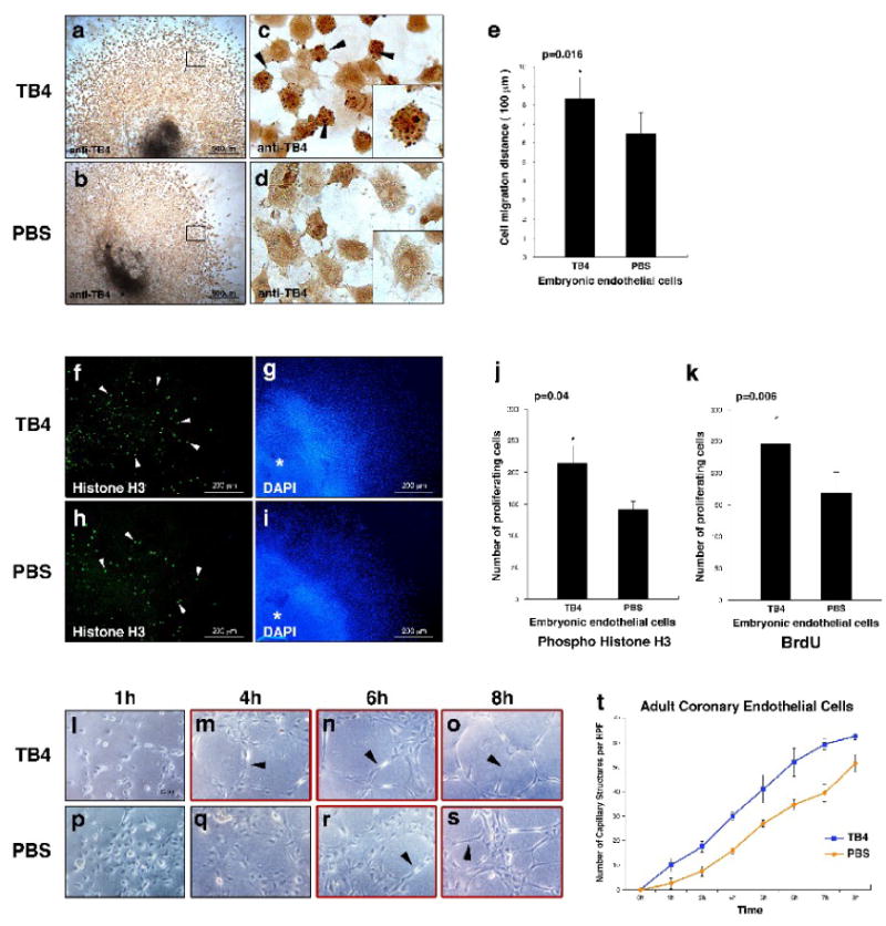
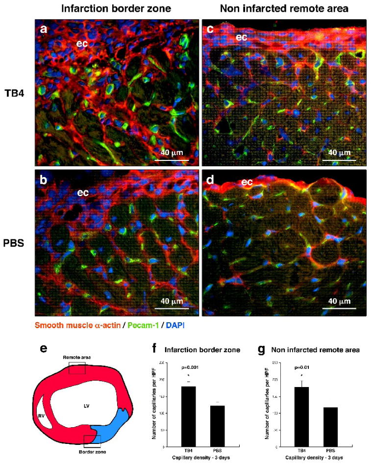
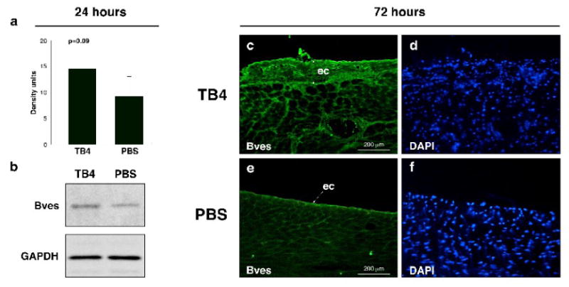
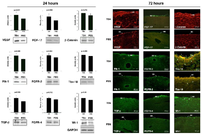
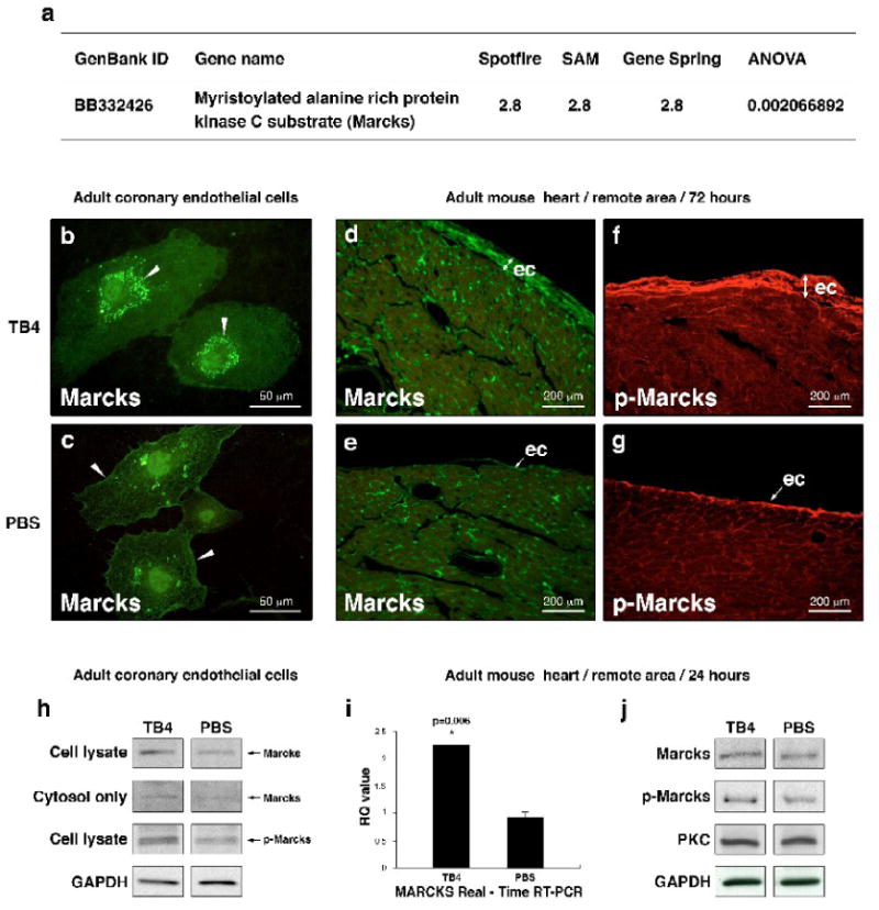
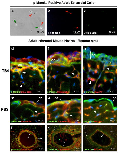
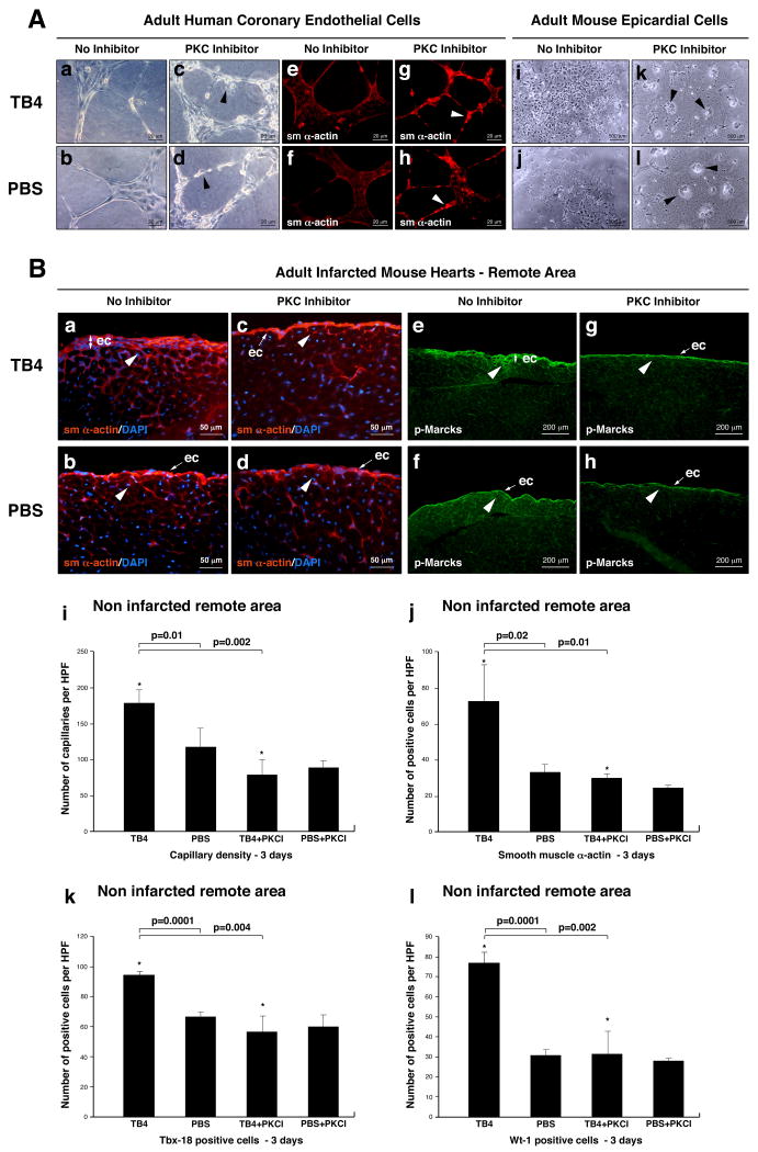
Hypoxic heart disease is a predominant cause of disability and death worldwide. Since adult mammalian hearts are incapable of regeneration after hypoxia, attempts to modify this deficiency are critical. As demonstrated in zebrafish, recall of the embryonic developmental program may be the key to success. Because thymosin beta4 (TB4) is beneficial for myocardial cell survival and essential for coronary development in embryos, we hypothesized that it reactivates the embryonic developmental program and initiates epicardial progenitor mobilization in adult mammals. We found that TB4 stimulates capillary-like tube formation of adult coronary endothelial cells and increases embryonic endothelial cell migration and proliferation in vitro. The increase of blood vessel/epicardial substance (Bves) expressing cells accompanied by elevated VEGF, Flk-1, TGF-beta, Fgfr-2, Fgfr-4, Fgf-17 and beta-Catenin expression and increase of Tbx-18 and Wt-1 positive myocardial progenitors suggested organ-wide recall of the embryonic program in the adult epicardium. TB4 also positively regulated the expression and phosphorylation of myristoylated alanine-rich C-kinase substrate (Marcks), a direct substrate and indicator of protein kinase C (PKC) activity in vitro and in vivo. PKC inhibition significantly reduced TB4 initiated epicardial thickening, capillary growth and the number of myocardial progenitors. Our results demonstrate that TB4 is the first known molecule capable of organ-wide activation of the embryonic coronary developmental program in the adult mammalian heart after systemic administration and that PKC plays a significant role in the process.


Figures

Figure 1
Figure 1. TB4 induces embryonic endothelial cell migration and proliferation and initiates capillary-like structure formation of HCECs in vitro
a–d, Mouse E11.5 cardiac outflow tract endothelial cells stained with anti-TB4/peroxidase after TB4 (a,c) or PBS (b,d) treatment. In c, black arrowheads and high magnification of a representative cell in the corner boxed area indicate increase in actively migrating cells with rounded morphology after TB4 treatment. (c) and (d) are high-magnification images of the boxed areas in (a) and (b). e, Distance of embryonic endothelial cell migration in E11.5 cardiac outflow tract explants with or without TB4 treatment. Bars indicate standard deviation at 95% confidence limits (n=6), *p < 0.05. f-k, Staining of mouse E11.5 cardiac outflow tract explants with phospho-histone H3 antibody (f,h) and 4,6-diamidino-2-phenylindole (DAPI) (g,i) shows increased endothelial cell proliferation (white arrowheads) after TB4 (f) treatment compared to PBS (h). Asterix indicates center of the explant. j-k, Number of proliferating embryonic endothelial cells with or without TB4 treatment. Bars indicate standard deviation at 95% confidence limits (n=6), *p < 0.05. l-s, Adult HCECs form capillary structures in advance when treated with TB4 (m-o black arrowheads) compared to PBS (r-s, black arrowheads) on matrigel. t, TB4 increases the number of HCEC capillary like structures on matrigel. Bars indicate standard deviation at 95% confidence limits (n=6).
Figure 2
Figure 2. TB4 initiates cardiac vessel formation in vivo
a-d, Immunohistochemistry using smooth muscle α-actin (red) and Pecam-1 (green) specific antibodies with DAPI staining (blue) revealed increase in capillary structures at the margin of the scar (a,b) and in the remote areas (c,d) three days after TB4 treatment (a,c) when compared to PBS (b,d). e, Schematic of adult mouse heart transverse sections used for histological analysis. Boxes indicate the areas investigated in (a-d). f-g, Number of capillaries increases significantly after TB4 treatment compared to PBS control. Bars indicate standard deviation at 95% confidence limits (n=6), *p < 0.05. LV, left ventricle; RV, right ventricle; ec, epicardium
Figure 3
Figure 3. Increase of Bves expression after TB4 treatment at the non infarcted remote areas of the adult mouse heart in vivo
a, Western blot analysis using Bves and GAPDH primary antibodies shows increase in Bves expression after TB4 treatment in the non infarcted cardiac tissue 24 h after ligation. b, Densitometric analysis of Western blot results (a) normalized to GAPDH loading control. Bars indicate standard deviation at 95% confidence limits (n=6). c-f, Immunohistochemical analysis with anti-Bves antibody shows increase in Bves-positive cells and organ-wide thickening of the remote epicardium 3 days after systemic TB4 treatment (c) compared to PBS (e). (d,f) are DAPI stain of (c,e). ec, epicardium
Figure 4
Figure 4. Systemic TB4 injection beneficially alters the expression of proteins essential for embryonic coronary development and myocardial progenitor activation in mammals and during cardiac regeneration in adult zebrafish
A. Densitometric analyses of Western blot results of adult cardiac tissue indicate significant changes in VEGF, Flk-1 and TGF-β expressions and moderate increase in FGF-17, FGFR-2, FGFR-4, β-Catenin, Tbx-18 and Wt-1 expressions 24 h after systemic TB4 injection when compared to PBS treatment in vivo. Heart tissue was harvested from the healthy, non infarcted areas of the hearts. Density units were normalized to GAPDH loading control. Bars indicate standard deviation at 95% confidence limits (n=6), *p < 0.05. B. Immunohistochemical analysis shows increase in VEGF, Flk-1, TGF-β, FGF-17, FGFR-2, FGFR-4, β-Catenin, Tbx-18 and Wt-1 expressions notably in the thickened epicardium at the intact areas of ligated adult mouse hearts 3 days after TB4 treatment when compared to PBS. White arrowheads in Tbx-18 sections indicate increase of Tbx-18 positive cells in TB4 treated epicardium and myocardium. White arrowheads in Wt-1 sections point at subepicardial localization of Wt-1 positive progenitors. ec, epicardium
Figure 5
Figure 5. TB4 alters Marcks expression in vitro and activates PKC in adult epicardium in vivo
a, cDNA microarray on adult mouse hearts reveals 2.8-fold increase in Marcks expression 24 h after TB4 treatment compared to PBS. i, Real-time RT-PCR of mouse heart RNA confirms the in vitro cDNA microarray results. Bars indicate standard deviation at 95% confidence limits (n=6), *p < 0.05. RQ, relative quantitation assay. b-c, Immunocytochemical analysis of HCECs with Marcks-specific antibody (green) shows increased Marcks expression and translocation from the cell membrane (c white arrowheads) to the cytosol (b white arrowheads) after TB4 treatment (b) suggesting a change in PKC activity by TB4 in vitro. h, Western blot of complete cell lysates and cytosol fractions from adult coronary endothelial cells 24 hours after TB4 or PBS treatment support translocation of Marcks protein into the cytosol and indicate increase in Marcks activation (p-Marcks) by PKC after TB4 treatment in vitro. d-g, Immunohistochemical analysis shows significant increase in Marcks (d,e) and phospho-Marcks (f,g) expressions in thickened epicardium at the intact areas of ligated adult mouse hearts 3 days after TB4 treatment (d,f)). j, Western blot analyses of adult cardiac tissue from the remote areas by Marcks, p-Marcks, PKC, and GAPDH antibodies 24 h after treatment show increase in Marcks expression and phosphorylation without significant change in PKC levels 24 hours after TB4 treatment in vivo (n=6).
Figure 6
Figure 6. p-Marcks labels future endothelial and smooth muscle cells in the adult epicardium
a-c, Immunocytochemical analysis of p-Marcks positive epicardial cells with sm α-actin (b) and p-Cytokeratin (c) primary antibodies indicate endothelial and/or smooth muscle cell fate seven days after culturing. (a) endothelial marker positive cells highlighted by green arrowheads, smooth muscle marker positive cells highlighted by red arrowheads. d-i, sm α-actin, Cytokeratin, Pecam-1 (red) and p-Marcks (green) co-immunostaining with DAPI (blue) reveals cellular heterogeneity in the TB4 activated (d,f,h) or in the single layered epicardium (e,g,i) of adult control hearts. White arrowheads in d-i indicate p-Marcks positive cells with endothelial or smooth muscle cell fate in the developing capillaries. j-l, Immunohistochemical analysis of the mature coronary vessels suggest smooth muscle and endothelial cell fate for p-Marcks positive cells. ec, epicardium; e, endothel; sm, smooth muscle; m, mesenchyme
Figure 7
Figure 7. PKC activity is essential for TB4 induced epicardial activation in adult mice in vivo
A, Bisindolylmaleinimide-I alters capillary formation of HCECs and inhibits adult epicardial cell transformation on matrigel. a-h, HCECs transform to irregular, rounded (c,d black arrowheads) sm α- actin positive cells (g,h white arrowheads) when treated with PKC inhibitor. i-l, Bisindolylmaleinimide-I inhibits adult epicardial cell transformation on matrigel (k,l black arrowheads point at non-transformed cell groups). B. Immunohistochemical analyses with anti-sm α-actin (a-d) and anti-p-Marcks (e-h) antibodies show reduced epicardial thickening and inhibition of capillary outgrowths (c,g white arrowheads) in TB4+PKC inhibitor treated adult hearts (c,g) when compared to TB4 and no inhibitor treated controls (d,h). Decrease in p-Marcks expression indicated sufficient reduction of PKC activity in the inhibited control hearts (h). Administration of Bisindolylmaleinimide-I itself does not alter the adult mouse epicardium in vivo (d,h). i-j, Bisindolylmaleinimide-I significantly suppresses the number of capillaries (i) and sm 28-actin positive cells (j) in the non infarcted remote areas of TB4 treated hearts (n=6). k-l, Inhibition of PKC activity significantly suppresses the number of TB4 activated Tbx-18 and Wt-1 positive myocardial progenitor cells in the non infarcted remote areas (n=6). Bars indicate standard deviation at 95% confidence limits. *p < 0.05. ec, epicardium

References

    1. Muñoz-Chápuli R, Quesada AR, Ángel Medina M. Angiogenesis and signal transduction in endothelial cells. Cell Mol Life Sci. 2004;61:2224–2243. - PMC - PubMed
    1. Carmeliet P. Mechanisms of angiogenesis and arteriogenesis. Nat Med. 2000;6:389–395. - PubMed
    1. Tomanek RJ. Formation of the coronary vasculature during development. Angiogenesis. 2005;8:273–284. - PubMed
    1. Mu H, Ohashi R, Lin P, Yao Q, Chen C. Cellular and molecular mechanisms of coronary vessel development. Vasc Med. 2005;10:37–44. - PubMed
    1. Beltrami AP, Barlucchi L, Torella D, Baker M, Limana F, Chimenti S, et al. Adult cardiac stem cells are multipotent and support myocardial regeneration. Cell. 2003;114:763–776. - PubMed

Publication types

MeSH terms