Review
. 2009 Apr;30(2):67-77.
doi: 10.1053/j.sult.2008.12.005.

Computed tomography and magnetic resonance imaging appearance of renal neoplasms after radiofrequency ablation and cryoablation

Affiliations
Review

Computed tomography and magnetic resonance imaging appearance of renal neoplasms after radiofrequency ablation and cryoablation

Satomi Kawamoto et al. Semin Ultrasound CT MR. 2009 Apr.

Abstract

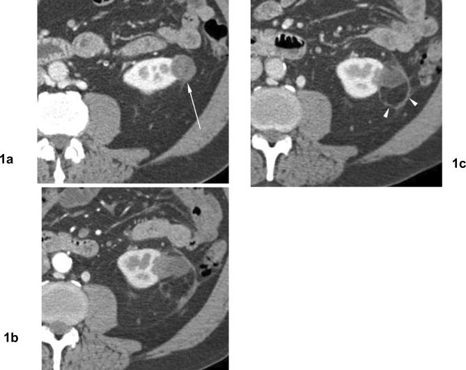
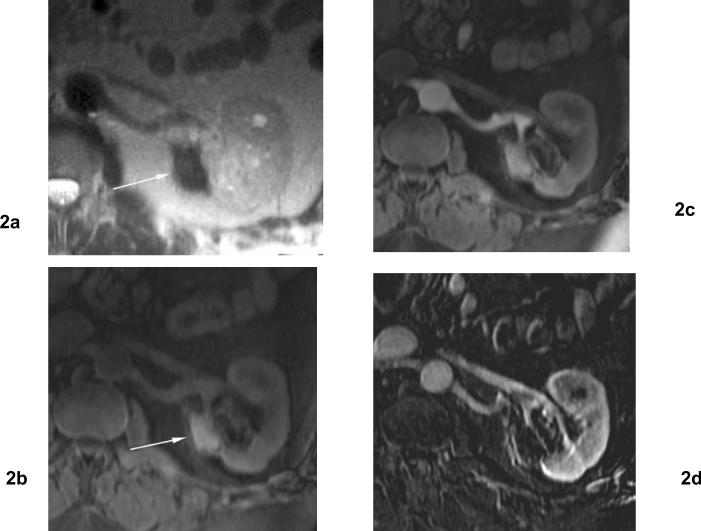
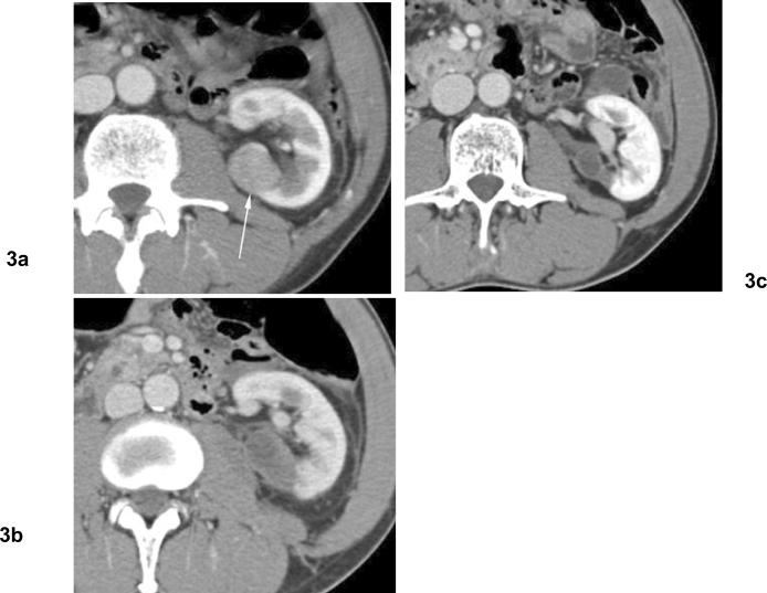
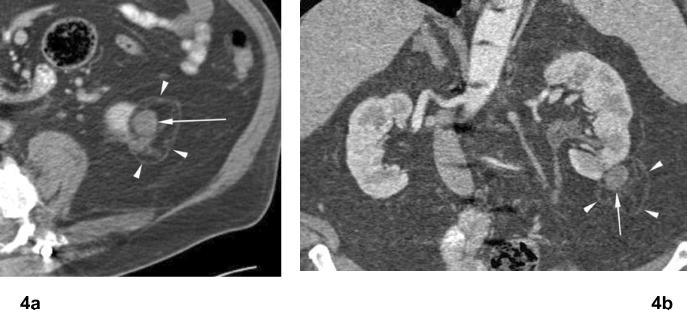
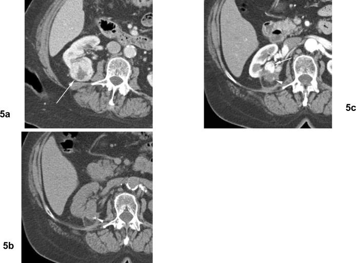
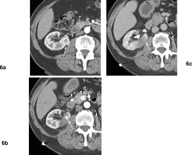
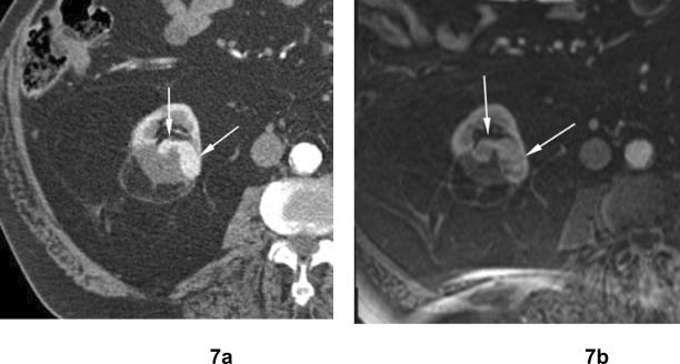
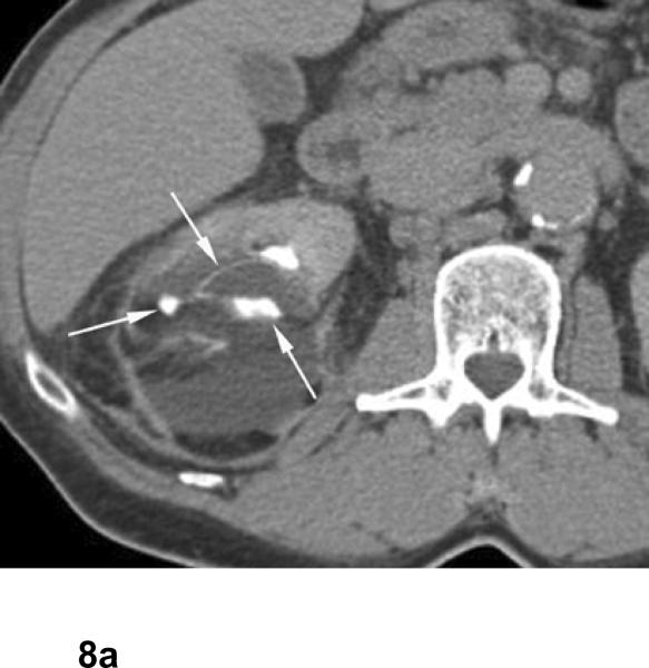
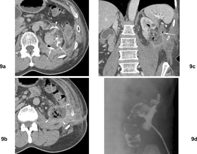
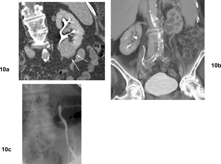
Thermal ablation procedures including radiofrequency ablation and cryoablation have been increasingly used for treatment for small renal cell carcinoma. Currently, computed tomography and magnetic resonance imaging are usually used to assess residual or recurrent disease after thermal ablation of renal tumor. After thermal ablation, the zone of ablation is usually seen as an area of hypoattenuation on computed tomography and is generally hypointense at T2-weighted magnetic resonance imaging and iso- to hyperintense at T1-weighted imaging relative to renal parenchyma. The ablation zone frequently involutes over time. Residual tumor after thermal ablation is most common at the margin of the ablation zone and often seen as nodular or crescent-shaped areas of contrast enhancement. Accurate assessment of ablated tumors at postprocedural imaging is essential for evaluating the adequacy of treatment and guiding further management. Complications are uncommon and usually minor but should be detected on postprocedural imaging studies.


Figures

Fig 1
Fig 1. Involution of treated tumor after cryoablation. Papillary type renal cell carcinoma
(a) Contrast enhanced axial CT before cryoablation shows exophytic hypodense mass in the left kidney (arrow). Biopsy showed papillary type renal cell carcinoma. (b) Venous phase axial CT 4 months after cryoablation shows heterogeneous hypodense area in the ablation zone with a curvilinear hyperattenuation area, or “halo”. (c) Venous phase axial CT 16 months after cryoablation shows decreasing in size of ablation zone without evidence of contrast enhancement. Persistent curvilinear hyperattenuation area in the perinephric fat surrounding the ablated tumor (arrowheads).
Fig 2
Fig 2. 4 months after cryoablation for clear cell type renal cell carcinoma of the left kidney
(a) Axial T2 weighted MR image shows a hypointense area with focal contour deformity (arrow) in the ablation zone. (b) Axial fat-suppressed T1 weighted MR image shows an area of hyperintensity in the ablation zone (arrow). (c) Axial contrast enhanced T1 weighted MR images obtained 4 months after cryoablation shows noncontrast enhancement in the ablation zone. (d) Subtraction image. The ablation zone demonstrates no evidence of enhancement.
Fig 3
Fig 3. Involution of treated tumor after cryoablation. Papillary type renal cell carcinoma
(a) Axial contrast enhanced CT before cryoablation shows solid mass in the left kidney posteriorly (arrow). Biopsy showed papillary type renal cell carcinoma. (b) Venous phase contrast enhanced CT 2 months after cryoablation shows heterogeneous hypodensity in ablation zone without contrast enhancement. (c) Venous phase contrast enhanced CT 10 months after cryoablation shows involution of treated tumor without contrast enhancement.
Fig 4
Fig 4. Treated tumor after RF ablation. Papillary type renal cell carcinoma
(a) Axial and (b) coronal contrast enhanced CT images 4 years after RF ablation shows persistent exophytic mass in lower pole of the left kidney, unchanged compared to early post ablation CT images (not shown). There is no contrast enhancement in ablated tumor (arrow). There is a curvilinear hyperattenuation area, or “halo” in perinephric fat surrounding ablated tumor (arrowheads).
Fig 5
Fig 5. Residual unablated tumor. Calcifications in ablation zone after cryoablation twice for clear cell type renal cell carcinoma
(a) Arterial phase contrast enhanced CT before cryoablation shows heterogeneously enhancing mass (arrow) in the right kidney posteriorly. (b) Noncontrast CT (15 months after 2nd cryoablation) shows small calcification (arrowhead) in the ablation zone. (c) Arterial phase contrast enhanced CT (15 months after 2nd cryoablation) shows nodular contrast enhancement at the periphery of ablation zone (arrow) indicating residual unablated tumor.
Fig 6
Fig 6. Residual unablated tumor after cryoablation. Clear cell type renal cell carcinoma. The patient is status post left nephrectomy for renal cell carcinoma
(a) Arterial phase contrast enhanced CT before cryoablation shows heterogeneously enhancing mass (arrow) in the right kidney medially. (b) Arterial phase contrast enhanced CT 1 month after cryoablation shows nodular contrast enhancement at the periphery of ablation zone (arrow) indicating residual unablated tumor. Note enhancing small masses in the pancreas (arrowheads) indicating metastatic foci. (c) Excretory phase contrast enhanced CT 1 months after cryoablation shows washout of contrast material from the nodular enhancement, and unablated tumor is seen as subtle hypodense area relative to normal renal parenchyma (arrow).
Fig 7
Fig 7. Residual unablated tumor after cryoablation. Clear cell type renal cell carcinoma
(a) Arterial phase contrast enhanced CT 6 months after cryoablation shows crescent-shaped areas of contrast enhancement (arrows) at the periphery of ablated zone in the right kidney indicating residual unablated tumor. (b) Axial contrast enhanced fat-suppressed T1-weighted MR image 12 months after cryoablation shows crescent-shaped areas of contrast enhancement in the right kidney which has slightly increased in size compared to (a).
Fig 8
Fig 8. Urinary leak from upper pole calyx after cryoablation for papillary renal cell carcinoma. The patient is 1 month after percutaneous cryoablation
Excretory phase axial and anterior volume rendered images show fluid collection around the ablated zone with contrast extravasation from upper pole calyx (arrows).
Fig 9
Fig 9. Left perinephric abscess and pyelocolonic fistula following open cryoablation of left renal cell carcinoma. Remote history of left partial nephrectomy for renal cell carcinomas
(a) Excretory phase axial CT 1 month after open cryoablation shows extravasation of contrast (arrows) in ablation zone. Ureter is displaced anteriorly (arrowhead) due to prior partial nephrectomy. (b) Excretory phase axial CT slightly more inferior to (a) shows an abscess inferior to the left kidney. (c) Venous phase coronal CT shows the abscess in ablation zone with air within the renal collecting system. The descending colon is closely related to this fluid collection (arrow). (d) Percutaneous drain was placed to the abscess, and urine leak was found in the draining fluid. Injection of contrast material shows communication of abscess to colon, indicating pyelocolonic fistula.
Fig 10
Fig 10. Ureteral stricture and hydronephrosis after cryoablation for clear cell type renal cell carcinoma
(a) Excretory phase coronal CT shows mass in the lower pole of the left kidney adjacent to the ureteropelvic junction. (b) Excretory phase coronal CT shows left hydronephrosis developed after cryoablation. (c) Retrograde ureterography shows obstruction of the left proximal ureter at the level of the ureteropelvic junction.

References

    1. Pantuck AJ, Zisman A, Belldegrun AS. The changing natural history of renal cell carcinoma. J Urol. 2001;166(5):1611–1623. - PubMed
    1. Chow WH, Devesa SS, Warren JL, Fraumeni JF., Jr. Rising incidence of renal cell cancer in the United States. Jama. 1999;281(17):1628–1631. - PubMed
    1. Jayson M, Sanders H. Increased incidence of serendipitously discovered renal cell carcinoma. Urology. 1998;51(2):203–205. - PubMed
    1. Luciani LG, Cestari R, Tallarigo C. Incidental renal cell carcinoma-age and stage characterization and clinical implications: study of 1092 patients (1982–1997) Urology. 2000;56(1):58–62. - PubMed
    1. Fergany AF, Hafez KS, Novick AC. Long-term results of nephron sparing surgery for localized renal cell carcinoma: 10-year followup. J Urol. 2000;163(2):442–445. - PubMed