Review
. 2009 Oct;11(10):2453-65.
doi: 10.1089/ars.2009.2558.

Mechanisms and function of DUOX in epithelia of the lung

Affiliations
Review

Mechanisms and function of DUOX in epithelia of the lung

Horst Fischer. Antioxid Redox Signal. 2009 Oct.

Abstract

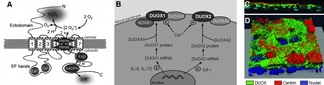
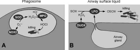
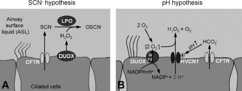
The human lung produces considerable amounts of H(2)O(2). In the normal uninflamed epithelium of both the airways and the alveoli, mucosal release of H(2)O(2) is readily detected both in cell cultures in vitro and in the exhaled breath of humans. The dual oxidases DUOX1 and DUOX2 are the H(2)O(2)-producing isoforms of the NADPH oxidase family found in epithelial cells. The DUOXs are prominently expressed at the apical cell pole of ciliated cells in the airways and in type II cells of the alveoli. Recent studies focused on the functional consequences of H(2)O(2) release by DUOX into the lung lining fluid. In the airways, a major function of DUOX is to support lactoperoxidase (LPO) to generate bactericidal OSCN(-), and there are indications that the DUOX/LPO defense system is critically dependent on the function of the CFTR Cl(-) channel, which provides both SCN(-) (for LPO function) and HCO(3)(-) (for pH adjustment) to the airway surface liquid. Although DUOX is also functional in the alveolar epithelium, no comparable heme peroxidase is present in the alveolus, and thus DUOX-mediated H(2)O(2) release by alveolar cells may have other functions, such as cellular signaling.


Figures

FIG. 1.
FIG. 1.
Overview of human airways. The proximal airways (trachea, bronchi, conducting bronchioles) are lined with a ciliated surface epithelium whose main surface cells are ciliated, goblet, and basal cells. The function of the proximal airways is to conduct gases and filter particles. There is ∼1 gland per square millimeter surface epithelium. The distal respiratory bronchioles are characterized by numerous alveoli, whose epithelium consists of type I and type II alveolar cells. The major function of the alveoli is gas exchange.
FIG. 2.
FIG. 2.
DUOX in the plasma membrane. (A) Model of DUOX in the membrane. The transmembrane domains are numbered 1 to 7. The NOX-typical gp91phox homology domain stretches from transmembrane domain 2 to the C terminus and contains the electron (e) transport chain, including binding sites for NADPH, FAD, and two hemes. O2•− forms as an intermediate and is internally dismutated to the final release product H2O2. DUOX releases cytosolic H+ during NADPH oxidation. Two intracellular EF hands bind Ca2+, and an ectodomain of unclear function might be involved in H2O2 metabolism. The products of DUOX function are extracellular H2O2 and intracellular H+. (B) Cellular regulation of DUOX1 and DUOX2 and currently identified interacting proteins. DUOX1 is specifically upregulated by IL-4 and IL-13 by ∼4-fold, while DUOX2 expression is highly induced by 20-fold by treatment of cells with interferon-γ [Inf-γ, (50)]. Both DUOXs interact with their respective ER-resident chaperone DUOXA1 and DUOXA2 (46), and the DUOXA proteins may be present and functional in the plasma membrane (76). In the apical membrane the function of the DUOXs is upregulated by intracellular Ca2+. NOXA1 has been shown to interact with and inhibit DUOXA1 in the plasma membrane in a Ca2+-dependent fashion (83). Circled + indicates stimulatory effect. (C and D) DUOX protein localizes to the apical pole of airways. Primary human airway epithelial culture was stained for DUOX (green), centrin (red), and nuclei (blue), as described (98). (C) Side view with apical aspect pointing up. DUOX is localized at the apical pole of ciliated, centrin-labeled cells. Nuclei at the bottom of the image are from basal cells, which do not stain for DUOX. (D) 3-D reconstructed confocal image stack at angled view onto the mucosa. Cells that do not stain for centrin (nonciliated cells, likely goblet cells) also do not stain for DUOX (Images in C and D by J. Tseng and H. Fischer, DUOX antibody kindly provided by F. Miot, centrin 20H5 antibody kindly provided by J.L. Salisbury.) (For interpretation of the references to color in this figure legend, the reader is referred to the web version of this article at www.liebertonline.com/ars).
FIG. 3.
FIG. 3.
Similar mechanisms of innate defense in phagocytes and the airways. (A) In phagocytic cells, the NOX2 complex produces O2•− inside the phagosome, which is dismutated to H2O2 by superoxide dismutase (SOD) and further reduced to bactericidal HOCl by myeloperoxidase (MPO). (B) In the airways, DUOX of the surface epithelium releases H2O2 into the airway surface liquid and lactoperoxidase (LPO), secreted mainly by submucosal glands (but also by surface goblet cells), produces bactericidal OSCN.
FIG. 4.
FIG. 4.
Proposed relations of CFTR to DUOX function. (A) SCN hypothesis. CFTR as an SCN conductor is required to supply the DUOX/LPO system with SCN for the conversion into bactericidal OSCN. Availability of SCN is the rate-limiting factor indicating that this system is inhibited in CF (17, 78). (B) pH hypothesis. CFTR as a HCO3 conductor is required to alkalinize the pH of the ASL to allow the HVCN1 H+ channel to release intracellular H+ produced by DUOX from NADPH. Stoichiometry is given for one NADPH oxidation cycle resulting in two intracellular H+ to generate one extracellular H2O2. Note, however, that HVCN1 does not discriminate between H+ of different intracellular origin and has been found to conduct a substantially higher H+ flux than expected from NADPH oxidation alone (98). Since both H+ and HCO3 are driven by the pH gradient, H+ flux only occurs into an alkaline ASL, while HCO3 is released into an acidic ASL (crossover point is approximately at pH6.9, H. Fischer, unpublished). Circled + indicates activation.

References

    1. Al–Obaidy A. Al–Samarai A. Exhaled breath condensate pH and hydrogen peroxide as non-invasive markers for asthma. Saudi Med J. 2007;28:1860–1863. - PubMed
    1. Ameziane–El-Hassani R. Morand S. Boucher J–L. Frapart Y–M. Apostolou D. Agnandji D. Gnidehou S. Ohayon R. Noel–Hudson M–S. Francon J. Lalaoui K. Virion A. Dupuy C. Dual oxidase-2 has an intrinsic Ca2+-dependent H2O2-generating activity. J Biol Chem. 2005;2280:30046–30054. - PubMed
    1. Antczak A. Kurmanowska Z. Kasielski M. Nowak D. Inhaled glucocorticosteroids decrease hydrogen peroxide level in expired air condensate in asthmatic patients. Respir Med. 2000;94:416–421. - PubMed
    1. Antczak A. Nowak D. Shariati B. Krol M. Piasecka G. Kurmanowska Z. Increased hydrogen peroxide and thiobarbituric acid-reactive products in expired breath condensate of asthmatic patients. Eur Respir J. 1997;10:1235–1241. - PubMed
    1. Ashino Y. Ying X. Dobbs LG. Bhattacharya J. [Ca2+]i oscillations regulate type II cell exocytosis in the pulmonary alveolus. Am J Physiol Lung Cell Mol Physiol. 2000;279:L5–13. - PubMed

Publication types

LinkOut - more resources