Review
. 2009 Aug;218(2):286-92.
doi: 10.1016/j.expneurol.2009.03.042. Epub 2009 Apr 7.

Amyloid beta, mitochondrial structural and functional dynamics in Alzheimer's disease

Affiliations
Review

Amyloid beta, mitochondrial structural and functional dynamics in Alzheimer's disease

P Hemachandra Reddy. Exp Neurol. 2009 Aug.

Abstract

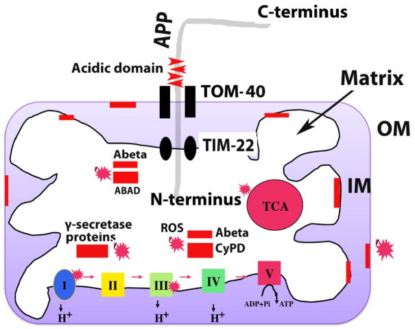
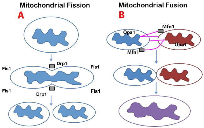
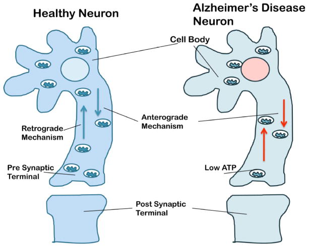
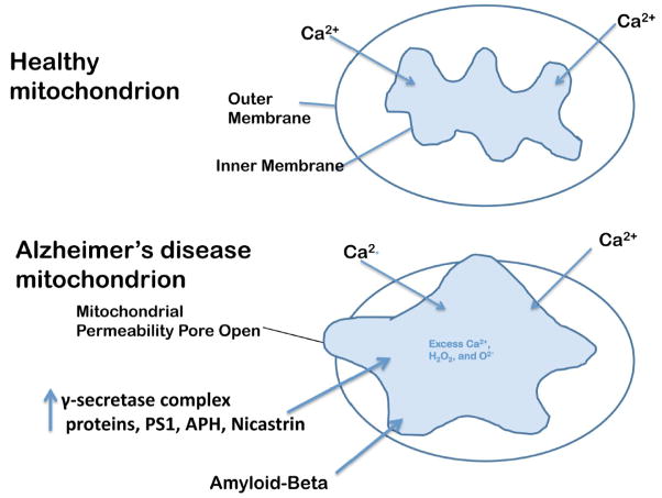
Mitochondria are the major source of energy for the normal functioning of brain cells. Increasing evidence suggests that the amyloid precursor protein (APP) and amyloid beta (Abeta) accumulate in mitochondrial membranes, cause mitochondrial structural and functional damage, and prevent neurons from functioning normally. Oligomeric Abeta is reported to induce intracellular Ca(2+) levels and to promote the excess accumulation of intracellular Ca(2+) into mitochondria, to induce the mitochondrial permeability transition pore to open, and to damage mitochondrial structure. Based on recent gene expression studies of APP transgenic mice and AD postmortem brains, and APP/Abeta and mitochondrial structural studies, we propose that the overexpression of APP and the increased production of Abeta may cause structural changes of mitochondria, including an increase in the production of defective mitochondria, a decrease in mitochondrial trafficking, and the alteration of mitochondrial dynamics in neurons affected by AD. This article discusses some critical issues of APP/Abeta associated with mitochondria, mitochondrial structural and functional damage, and abnormal intracellular calcium regulation in neurons from AD patients. This article also discusses the link between Abeta and impaired mitochondrial dynamics in AD.


Figures

Figure 1
Figure 1
APP and Ab localization of mitochondria in neurons from patients with AD. APP is localized to mitochondria only in AD patients, and APP localization increases with disease severity of Alzheimer’s. As shown, the N-terminus of APP is localized to mitochondria and the C-terminus, facing out. Mitochondrially localized APP forms complexes with the translocase of the outer membrane (TOM) and of the inner membrane (TIM). However, the transmembrane-arrested form of APP (ACID domain) prevents the importation of the C-terminus of APP into mitochondria, and the mitochondrial pores are clogged by APP import and prevent (i.e., block) the transport of nuclear-encoded mitochondrial proteins to the mitochondria. Insufficient nuclear-encoded mitochondrial proteins transported to the mitochondria and cause abnormal mitochondrial electron activities, including increased free radical production, decreased mitochondrial enzyme activities, and low ATP production. Ab was found in the outer and inner mitochondrial membranes and matrix. Ab associated localized to the outer membrane blocks the transport of nuclear-encoded mitochondrial proteins to the mitochondria and impairs ETC activities and ATP production in AD neurons. Ab localized to the inner membrane directly induced free radical production, increased free radical production, decreased mitochondrial enzyme activities including cytochrome oxidase activity, and interfered with ATP production. Ab localized to the mitochondrial matrix interacted with several matrix proteins, including ABAD and Cyclophilin D. The abnormal interaction between Ab and mitochondrial proteins caused mitochondrial damage and promoted neuronal damage in AD patients. Further, g-secretase complex proteins, PS1, APH and nicastrin were found in the matrix of mitochondria and may damage mitochondria by inducing free radical production and causing oxidative damage to mitochondria.
Figure 2
Figure 2
Mitochondrial fission and fusion events in neurons. This figure shows the mitochondrial shape and structure are maintained by 2 opposing forces: mitochondrial fission (A) and mitochondrial fission (B). In a healthy neuron, fission and fusion mechanisms balance equally. Mitochondria alter their shape and size to move from cell body to the axons, dendrites, and synapses, and back to the cell body through mitochondrial trafficking. Fission and fusion are controlled by evolutionary conserved, large GTPases belonging to the family of dynamin. Fission is controlled and regulated by dynamin related protein (Drp1) and fission (Fis1); Fis1 is localized to the outer membrane of mitochondria. Most of the Drp1 protein is localized in the cytoplasm but a small part punctures the outer membrane, which promotes the fragmentation of mitochondria. Increased mitochondrial free radicals activate Fis1, which is also critical for mitochondrial fission. Mitochondrial fusion is controlled by 3 GTPase proteins: 2 outer-membrane localized proteins Mfn1 and Mfn2, and 1 inner-membrane localized protein Opa1. The C-terminal part of Mfn1 mediates oligomerization between Mfn molecules of adjacent mitochondria and facilitates the fusing of mitochondria. In AD neurons, mitochondrially generated free radicals activate Fis1 and promote increased mitochondrial fragmentation; this increased mitochondrial fragmentation produces defective mitochondria that ultimately damage neurons.
Figure 3
Figure 3
Mitochondrial trafficking in neurons from healthy subjects and patients with AD. Mitochondria are synthesized in the cell body and travel along the axons and dendrites to supply energy to nerve terminals for normal neural communication; they travel back to the cell body via mitochondrial trafficking. Mitochondria are transported from the cell body to nerve terminals via an anterograde mechanism and from nerve terminals to the cell body via a retrograde mechanism. In healthy and functionally active neurons, anterograde and retrograde transport of mitochondria are equal and active. In AD neurons, both anterograde and retrograde transport of mitochondria are slow because of the presence of large number of defective and functionally inactive mitochondria. These defective mitochondria are not able to supply sufficient levels of energy at nerve terminals, which may impair neurotransmission, and ultimately result in synaptic damage, neurodegeneration, and cognitive decline in AD patients.
Figure 4
Figure 4
Opening of the mitochondrial permeability transition pore. The increased entry of APP and Ab into mitochondria, and the increase in Ca2+ levels in the matrix of mitochondria may cause the inner mitochondrial pore to open. In general, the inner mitochondrial membrane provides a highly efficient barrier to ionic flow and protects mitochondria from toxic insults. However, in neurons from patients with AD, an age-dependent accumulation of APP and Ab1–42 oligomers and g-secretase complex proteins, PS1, APH and nicastrin may induce a massive entry of Ca2+ into neurons and may promote mitochondrial Ca2+ overload. Excess mitochondrial Ca2+ may promote the opening of the mitochondrial permeability transition pore and may destroy the neuron by apoptotic cell death.

References

    1. Almeida CG, Tampellini D, Takahashi RH, Greengard P, Lin MT, Snyder EM, Gouras GK. Beta-amyloid accumulation in APP mutant neurons reduces PSD-95 and GluR1 in synapses. Neurobiol Dis. 2005;20:187–198. - PubMed
    1. Anandatheerthavarada HK, Biswas G, Robin MA, Avadhani NG. Mitochondrial targeting and a novel transmembrane arrest of Alzheimer's amyloid precursor protein impairs mitochondrial function in neuronal cells. J Cell Biol. 2003;161:41–54. - PMC - PubMed
    1. Anandatheerthavarada HK, Devi L. Amyloid precursor protein and mitochondrial dysfunction in Alzheimer’s disease. Neuroscientist. 2007 Dec;13:626–638. - PubMed
    1. Baloyannis SJ, Mauroudis I, Manolides SL, Manolides LS. Synaptic alterations in the medial geniculate bodies and the inferior colliculi in Alzheimer's disease: a Golgi and electron microscope study. Acta Otolaryngol. 2008;30:1–3. - PubMed
    1. Benard G, Bellance N, James D, Parrone P, Fernandez H, Letellier T. Rossignol R.Mitochondrial bioenergetics and structural network organization. J Cell Sci. 2007;120:838–848. - PubMed

Publication types

Substances