. 2009 Jun;45(9):1709-20.
doi: 10.1016/j.ejca.2009.03.013. Epub 2009 Apr 7.

Growth inhibitory and anti-tumour activities of OSU-03012, a novel PDK-1 inhibitor, on vestibular schwannoma and malignant schwannoma cells

Affiliations

Growth inhibitory and anti-tumour activities of OSU-03012, a novel PDK-1 inhibitor, on vestibular schwannoma and malignant schwannoma cells

Tina X Lee et al. Eur J Cancer. 2009 Jun.

Abstract

Background: Vestibular schwannomas (VS) frequently express high levels of activated AKT. Small-molecule inhibitors of AKT signalling may have therapeutic potential in suppressing the growth of benign VS and malignant schwannomas.

Method: Primary VS and Schwann cells, human malignant schwannoma HMS-97 cells and mouse Nf2(-/-) Schwann cells and schwannoma cells were prepared to investigate the growth inhibitory and anti-tumour activities of OSU-03012, a celecoxib-derived small-molecule inhibitor of phosphoinositide-dependent kinase-1. Cell proliferation assays, apoptosis, Western blot, in vivo xenograft analysis using SCID mice and immunohistochemistry were performed.

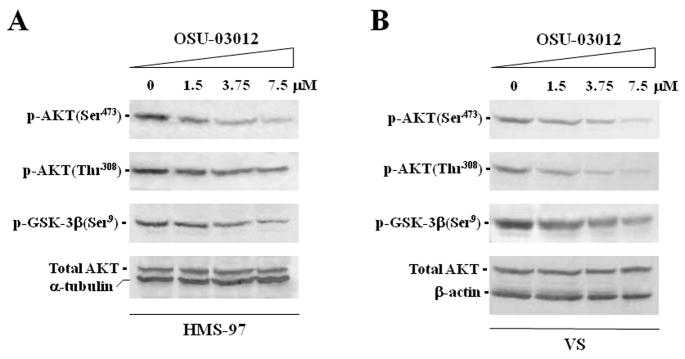
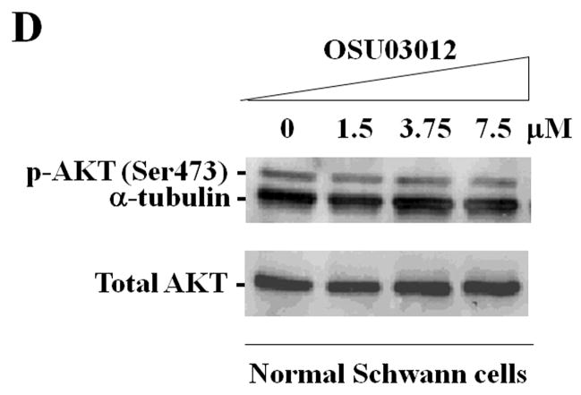
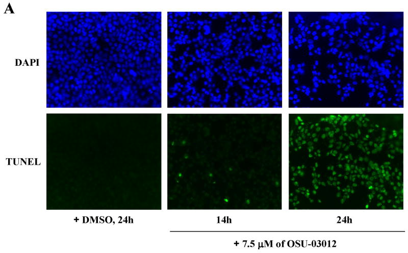
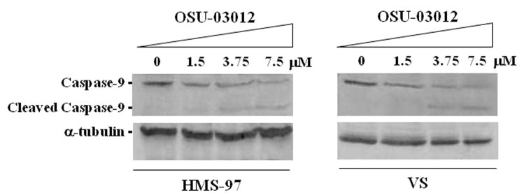
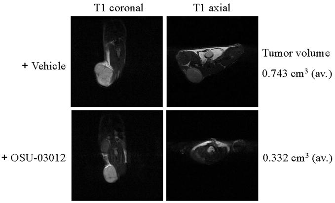
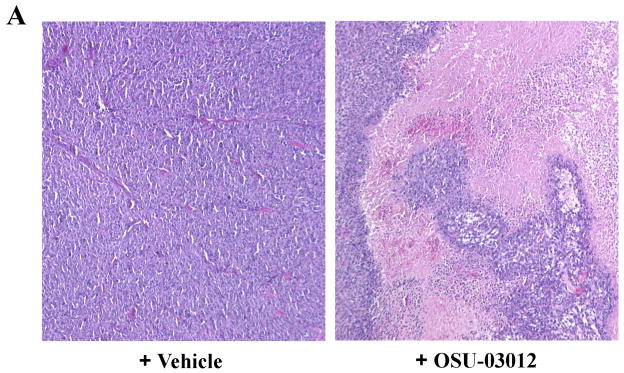
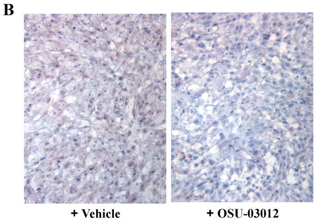
Results: OSU-03012 inhibited cell proliferation more effectively in both VS and HMS-97 cells than in normal human Schwann cells. The IC5) of OSU-03012 at 48h was approximately 3.1 microM for VS cells and 2.6 microM for HMS-97 cells, compared with the IC(50) of greater than 12 microM for human Schwann cells. Similarly, mouse Nf2(-/-) schwannoma and Nf2(-/-) Schwann cells were more sensitive to growth inhibition by OSU-03012 than wild-type mouse Schwann cells and mouse schwannoma cells established from transgenic mice carrying the NF2 promoter-driven SV40 T-antigen gene. Like VS cells, malignant schwannoma HMS-97 cells expressed high levels of activated AKT. OSU-03012 induced apoptosis in both VS and HMS-97 cells and caused a marked reduction of AKT phosphorylation at both the Ser-308 and Thr-473 sites in a dose-dependent manner. In vivo xenograft analysis showed that OSU-03012 was well tolerated and inhibited the growth of HMS-97 schwannoma xenografts by 55% after 9 weeks of oral treatment. The anti-tumour activity correlated with reduced AKT phosphorylation.

Conclusion: OSU-03012 is a potential chemotherapeutic agent for VS and malignant schwannomas.


Conflict of interest statement

Conflict of interest statement

All authors do not have any disclosure of potential conflict of interest.

Figures

Figure 1
Figure 1. Effects of OSU-03012 on the proliferation of human VS cells grown in the absence (A) or presence (B) of heregulin, human malignant schwannoma HMS-97 cells (C), and normal human Schwann cells (D)
Primary human VS and Schwann cells were prepared as described in Materials and Methods, and were plated out at a density of 5 × 10³ cells/well in a poly-D-lysine/laminin-coated 96-well plate in growth medium with or without heregulin at 37°C for 24 hours. HMS-97 cells were seeded at the same density in DME medium supplemented with 10% FBS overnight. Various concentrations of OSU-03012 were then added to the culture. DMSO (0.05%) in which the stock solution of OSU-03012 was prepared was used as the vehicle control. After treatment for 48 hours, cell proliferation was measured using the CellTiter 96® AQueous One Solution Cell Proliferation Assay (Promega) and recorded using a Victor 1420 Multilabel Counter (Perkin Elmer). The percentage of cell proliferation was plotted against the concentrations of OSU-03012. The experiments were performed in six replicates. Columns show the mean of the replicates and error bars denote the standard deviation. Six different preparations of primary VS cells (Table 1) were studied and the representative result was shown.
Figure 2
Figure 2. Effects of OSU-03012 on the proliferation of (A) Nf2−/− mouse schwannoma cells from the P0Cre;Nf2flox/flox mouse, (B) Nf2−/− mouse Schwann cells, (C) Nf2+/+ mouse Schwann cells, and (D) T antigen-transformed mouse NF2P2.4-T schwannoma cells
Various mouse schwannoma and Schwann cells were plated out in a poly-D-lysine/laminin-coated 96-well plate (5 × 10³ cells/well) in complete growth medium at 37°C for 24 hours and then treated with various concentrations of OSU-03012 or DMSO control for another 48 hours before cell proliferation assay was performed. The percentage of cell proliferation was plotted against the concentrations of OSU-03012. Each experiment was performed in triplicates and was repeated at least two times. Columns show the mean of the replicates and error bars denote the standard deviation.
Figure 3
Figure 3. Effects of OSU-03012 on AKT phosphorylation in human malignant schwannoma HMS-97 cells (A), primary human VS cells (B), mouse NF2P2.4-T schwannoma cells (C), and normal mouse Schwann cells (D)
Various concentrations of OSU-03012 were added to the cells for 12 hours. Cell lysates were analysed for p-AKT(Ser473), p-AKT(Thr308), p-GSK-3β (Ser 9), and total AKT by Western blots. In addition, the expression of α-tubulin or β-actin was detected and served as a loading control.
Figure 4
Figure 4. OSU-03012 treatment induced apoptosis in both human malignant schwannoma HMS-97 cells (A) and primary human VS cells (B)
OSU-03012 (7.5 μM) were added to the cells for the indicated time periods. As controls, HMS-97 cells were treated with DMSO (0.05%) for 24 hours and VS cells were treated for 48 hours. Treated cells were fixed and labeled by TUNEL staining (shown in green). DAPI was used to counter stain the nuclei in blue.
Figure 5
Figure 5. OSU-03012 treatment induced cleavage of caspase 9
Human malignant schwannoma HMS-97 cells and primary human VS cells were treated with increasing concentrations of OSU-03012 for 12 hours. Soluble cell lysates were prepared and analysed by Western blot using an anti-caspase-9 antibody to detect pro-caspase-9 (47 kD) and the cleaved caspase 9 fragment (37 kD). The expression of α-tubulin was also detected and used as a loading control.
Figure 6
Figure 6. Volumetric measurement of human malignant schwannoma xenografts in SCID mice with or without OSU-03012 treatment by MRI
HMS-97 cells (2.5 × 10⁵ cells/mouse) were implanted in the right flank of SCID mice as previously described. The mice were observed for one week to allow the growth of xenografts. Subsequently, one group of mice was administered 200 mg/kg/day of OSU-03012 by oral gavage. The other group of mice was gavages-fed a similar volume of the vehicle as the control. After nine weeks of treatment, mice were examined under anesthesia using a 4.7 Tesla/cm MRI scanner (Bruker). Both T1-weighted axial and coronal images were obtained and multi-planar tumour volumes were determined as described in Materials and Methods. Images from representative mice are shown. Note that the average tumour volume of schwannoma xenografts was greatly reduced in the OSU-03012-treated group.
Figure 7
Figure 7. Immuno-histopathological examination of HMS-97 xenograft tumours from SCID mice with or without OSU-03012 treatment
(A) Hematoxylin-eosin staining of tissue sections of HMS-97 tumours from mice treated with OSU-03012 or vehicle control for nine weeks by oral gavages. (B) Immunostaining for p-AKT(Ser473) expression in HMS-97 tumour tissue sections from mice treated with OSU-03012 or vehicle control for nine weeks.

References

    1. Welling DB, Packer MD, Akhmametyeva EM, Chang LS. The Biology and Genetics of Vestibular Schwannomas. In: Bambakidis NC, Megerian, Porter, Spetzler RF, editors. Tumors of the Cerebellopontine Angle. B.C. Decker, Inc; 2007.
    1. Rouleau GA, Merel P, Lutchman M, et al. Alteration in a new gene encoding a putative membrane-organising protein causes neurofibromatosis type 2. Nature. 1993;363:515–521. - PubMed
    1. Trofatter JA, MacCollin MM, Rutter JL, et al. A novel Moesin-, Exrin-, Radixin-like gene is a candidate for the neurofibromatosis 2 tumor-suppressor. Cell. 1993;72:791–800. - PubMed
    1. Welling DB, Lasak JM, Akhmametyeva E, Ghaheri B, Chang LS. cDNA microarray analysis of vestibular schwannomas. Otol Neurotol. 2002;23:736–748. - PubMed
    1. Shin M, Ueki K, Kurita H, Kirino T. Malignant transformation of a vestibular schwannoma after gamma knife radiosurgery. Lancet. 2002;360:309–310. - PubMed

Publication types

MeSH terms