. 2009 Jun 5;284(23):15880-93.
doi: 10.1074/jbc.M109.005694. Epub 2009 Apr 9.

Discovery and exploitation of inhibitor-resistant aurora and polo kinase mutants for the analysis of mitotic networks

Affiliations

Discovery and exploitation of inhibitor-resistant aurora and polo kinase mutants for the analysis of mitotic networks

Paul J Scutt et al. J Biol Chem. .

Abstract

The Aurora and Polo-like kinases are central components of mitotic signaling pathways, and recent evidence suggests that substantial cross-talk exists between Aurora A and Plk1. In addition to their validation as novel anticancer agents, small molecule kinase inhibitors are increasingly important tools to help dissect clinically relevant protein phosphorylation networks. However, one major problem associated with kinase inhibitors is their promiscuity toward "off-target" members of the kinome, which makes interpretation of data obtained from complex cellular systems challenging. Additionally, the emergence of inhibitor resistance in patients makes it clear that an understanding of resistance mechanisms is essential to inform drug design. In this study, we exploited structural knowledge of the binding modes of VX-680, an Aurora kinase inhibitor, and BI 2536, a Polo-like kinase inhibitor, to design and evaluate drug-resistant kinase mutants. Using inducible stable human cell lines, we authenticated mitotic targets for both compounds and demonstrated that Aurora A mutants exhibit differential cellular sensitivity toward the inhibitors VX-680 and MLN8054. In addition, we validated Aurora B as an important anti-proliferative target for VX-680 in model human cancer cells. Finally, this chemical genetic approach allowed us to prove that Aurora A activation loop phosphorylation is controlled by a Plk1-mediated pathway in human cells.


Figures

FIGURE 1.
FIGURE 1.
Biochemical analysis of drug-resistant Aurora A mutants. A, the kinase catalytic subdomain V sequences are aligned. The gatekeeper residue is shown in red, and the positions of the Gly residues found in VX-680-sensitive kinases is indicated. h, human; x, Xenopus. B, Aurora A inhibition was assessed in duplicate. Activity is reported as a percentage of the control calculated from incubations containing 2.5% (v/v) DMSO. IC50 values reported represent the means determined from two or more independent experiments. Aurora A phosphorylation at Thr288 (pT288 AurA) was determined by immunoblotting. Equal loading was demonstrated with a pan-Aurora A antibody (AurA). C, WT and G216L Aurora A proteins were assayed in the presence of VX-680. Proteins were separated by SDS-PAGE, and phosphate incorporation was visualized by autoradiography. D, shown is the complex of Aurora A and VX-680, highlighting the drug-binding site adjacent to Gly216 (left) and modeled with Leu in the G216L mutant (right). E, activation and inhibition of WT, G216L, or D274A Aurora A was assessed in the presence of GST or GST-TPX2 and 300 nm VX-680. GST-TPX2 stained particularly strongly with Coomassie dye compared with GST. F and G, inhibition of WT or G216L Aurora A was assessed in the presence of the indicated concentrations of ZM447439 and MLN8054, respectively. Control incubations contained 2.5% (v/v) DMSO. Calculated IC50 values represent the means determined from two independent experiments.
FIGURE 2.
FIGURE 2.
Inhibition of recombinant Plk1 enzymes. A, shown is the inhibition of Sf9-expressed human Plk1 by BI 2536. Full-length His-Plk1 was assayed in duplicate in the presence of 100 μm [γ-32P]ATP and the indicated concentrations of BI 2536 using α-casein (red line) or Plk1 (black line) as substrate. Activity is reported as a percentage of the control calculated from incubations containing 2.5% (v/v) DMSO. B, shown is the complex of Plk1 and BI 2536, highlighting residues adjacent to the drug-binding site, including Arg136 (left), and as modeled with Gly in the R136G mutant (right). C, T210D Plk1 and R136G/T210D Plk1 catalytic domains were assayed in the presence of 100 μm [γ-32P]ATP and the indicated concentrations of BI 2536 (left). Equal Plk1 loading was demonstrated using horseradish peroxidase-conjugated anti-His antibody. Phosphorylated casein bands were quantified, and the data are plotted graphically (right). Activity is reported as a percentage of the control calculated from incubations containing 2.5% (v/v) DMSO. Similar results were seen in two independent experiments. D, conditions were the same as described for C, except that the unrelated Plk inhibitor GW843682X replaced BI 2536.
FIGURE 3.
FIGURE 3.
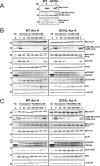
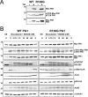
Analysis of HeLa cells harboring inducible Aurora A genes. A, WT or G216L human Aurora A-encoding Myc-tagged plasmids were stably integrated in HeLa cells, with expression being dependent on the addition of Tet. Cell extracts were analyzed by Western blotting with antibodies recognizing Myc (upper), phospho-Thr288 Aurora A (pT288 AurA; middle), and total Aurora A (AurA; lower). B and C, Tet-exposed asynchronous (AS) and mitotic cells expressing WT or G216L Aurora A were plated for 2 h in the presence or absence of the indicated concentrations of VX-680 and MLN8054, respectively. Cell lysates were probed with the indicated antibodies. Similar results were seen in two independent experiments.
FIGURE 4.
FIGURE 4.
Analysis of human DLD-1 cells harboring inducible Aurora A genes. WT or G216L human Aurora A-encoding Myc-tagged plasmids were stably integrated in Tet-inducible DLD-1 cells. Asynchronous cells were incubated overnight in the absence or presence of Tet prior to treatment with 20 μm MG132 and DMSO or 200 nm VX-680 for 2 h. Cells were subsequently fixed in methanol prior to processing for immunofluorescence. WT (A) or G216L (B) Aurora A (Aur A)-expressing cells were co-stained with antibodies to Myc (red), phospho-Thr288 Aurora A (pT288 AurA; green), total Aurora A (AurA), and 4′,6-diamidino-2-phenylindole (DAPI; blue). Merged color plots are shown to demonstrate co-localization of proteins. Monopolar (C) or misaligned (D) chromosome phenotypes were scored based on Aurora A fluorescence and DNA staining and morphology in the presence or absence of Tet and 200 nm VX-680. The graphs show the mean ± S.E. (95% confidence interval) from two experiments (>50 individual cells). Comparable results were obtained in several independent experiments.
FIGURE 5.
FIGURE 5.
Analysis of HeLa cells harboring inducible Aurora B genes. A, WT or G160L human Aurora B-encoding Myc-tagged plasmids were stably integrated in HeLa cells. Cell extracts were analyzed by Western blotting with antibodies recognizing Myc (upper) or Aurora B (AurB; lower). B, Tet-exposed asynchronous (AS) and mitotic cells expressing WT or G160L Aurora B were cultured in the presence or absence of the indicated concentrations of VX-680. Cell lysates were probed with the indicated antibodies. Similar results were seen in two independent experiments. C, asynchronous WT Aurora B HeLa cells (upper) or G160L Aurora B cells (lower) were incubated with or without Tet, fixed in methanol, and stained with the indicated antibodies. Note that the anti-phospho-Thr232 Aurora B antibody (pT232 AurB) also cross-reacted with phospho-Thr288 Aurora A on the centrosome. D, spindle morphology was scored in cell lines exposed to VX-680 in the presence and absence of Tet. E, chromosome alignment defects were scored in cells exposed to VX-680 in the presence and absence of Tet. Graphs show the mean ± S.E. (95% confidence interval) from two experiments.
FIGURE 6.
FIGURE 6.
Effects of VX-680 in HeLa cells overexpressing resistant Aurora A and B mutants. The DNA content of asynchronous HeLa cells overexpressing WT or G216L Aurora A (A) or WT or G160L Aurora B (B) was analyzed by flow cytometry. Cells were treated with DMSO or 50 nm VX-680 for 24 h prior to fixation staining. The Western blots after 7 days of continuous culture in Tet are shown (C). Cell extracts were incubated with anti-Myc (upper) or anti-total Aurora A (AurA) or B (AurB) (lower) antibodies. Cells were plated at low density as described under “Experimental Procedures.” Tet was added overnight prior to the addition of inhibitor and was reapplied on the 3rd day. Each well was treated with the indicated concentrations of VX-680 (D) or MLN8054 (E) on day 1 of the experiment. The cloning efficiency of the DMSO controls was set at 100%, and quantified data for the Tet-induced cells are indicated. Similar results were seen in two independent duplicate experiments.
FIGURE 7.
FIGURE 7.
Analysis of Plk1-expressing cell lines. A, WT or R136G human Plk1-encoding Myc-tagged plasmids were stably integrated in HeLa cells. Cell extracts were analyzed by Western blotting with antibodies recognizing Myc (upper), phospho-Thr210 Plk1 (pT210 Plk1; middle) or total Plk1 (lower). B, Tet-exposed asynchronous (AS) and mitotic cells expressing WT or R136G Plk1 were incubated for 2 h in the presence of the proteasome inhibitor MG132 (20 μm) and in the presence or absence of the indicated concentrations of BI 2536. Cell lysates were probed with the indicated antibodies, and anti-α-tubulin antibody was used to demonstrate equal protein loading in each lane. Similar results were seen in several independent experiments. AurA, Aurora A.
FIGURE 8.
FIGURE 8.
Expression of WT Plk1 and BI 2536-resistant Plk1. Tet-regulated WT Plk1 (A) and R136G Plk1 (B) were stably expressed in HeLa cells. Cells were treated overnight in the absence or presence of Tet and then fixed in methanol prior to processing for immunofluorescence. Representative images of mitotic cells in telophase depict Myc (red), Plk1 (green), and total Aurora A and 4′,6-diamidino-2-phenylindole (DAPI; blue). The merged color plots demonstrate co-localization of proteins. An asynchronous cell population expressing WT Plk1 (C) or R136G Plk1 (D) was treated with 20 μm MG132 and DMSO or 20 nm BI 2536 for 2 h and then processed for immunofluorescence. Cells were stained with antibodies to Myc, phospho-Thr288 Aurora A (pT288 AurA; green), total Aurora A (AurA; red), and 4′,6-diamidino-2-phenylindole (DAPI; blue). A monopolar phenotype was scored and quantified based on anti-Aurora A antibody fluorescence and DNA morphology (n = >50 cells) (E). The graph shows the mean ± S.E. (95% confidence interval) from two experiments. Similar results were observed in two independent experiments.

References

    1. Cohen P. ( 2009) Curr. Opin. Cell Biol. 21, 317– 324 - PubMed
    1. Zhang J., Yang P. L., Gray N. S. ( 2009) Nat. Rev. Cancer 9, 28– 39 - PubMed
    1. Druker B. J. ( 2008) Blood 112, 4808– 4817 - PubMed
    1. Fabian M. A., Biggs W. H., 3rd, Treiber D. K., Atteridge C. E., Azimioara M. D., Benedetti M. G., Carter T. A., Ciceri P., Edeen P. T., Floyd M., Ford J. M., Galvin M., Gerlach J. L., Grotzfeld R. M., Herrgard S., Insko D. E., Insko M. A., Lai A. G., Lélias J. M., Mehta S. A., Milanov Z. V., Velasco A. M., Wodicka L. M., Patel H. K., Zarrinkar P. P., Lockhart D. J. ( 2005) Nat. Biotechnol. 23, 329– 336 - PubMed
    1. Bain J., Plater L., Elliott M., Shpiro N., Hastie C. J., McLauchlan H., Klevernic I., Arthur J. S., Alessi D. R., Cohen P. ( 2007) Biochem. J. 408, 297– 315 - PMC - PubMed

Publication types

LinkOut - more resources