. 2009 Jun 12;284(24):16082-16089.
doi: 10.1074/jbc.M809282200. Epub 2009 Apr 9.

Ubx4 modulates cdc48 activity and influences degradation of misfolded proteins of the endoplasmic reticulum

Affiliations

Ubx4 modulates cdc48 activity and influences degradation of misfolded proteins of the endoplasmic reticulum

Sven M Alberts et al. J Biol Chem. .

Abstract

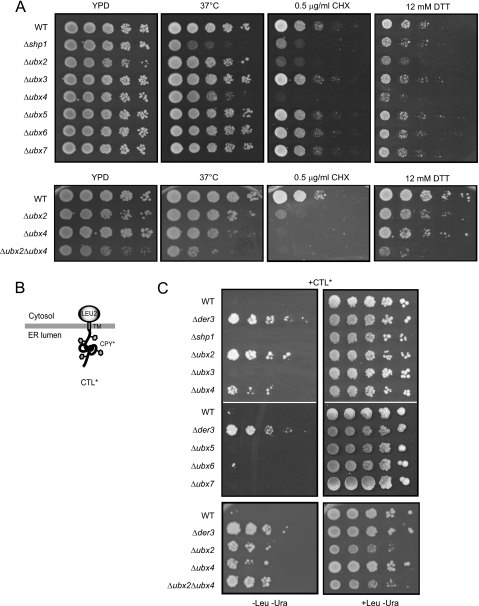
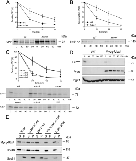
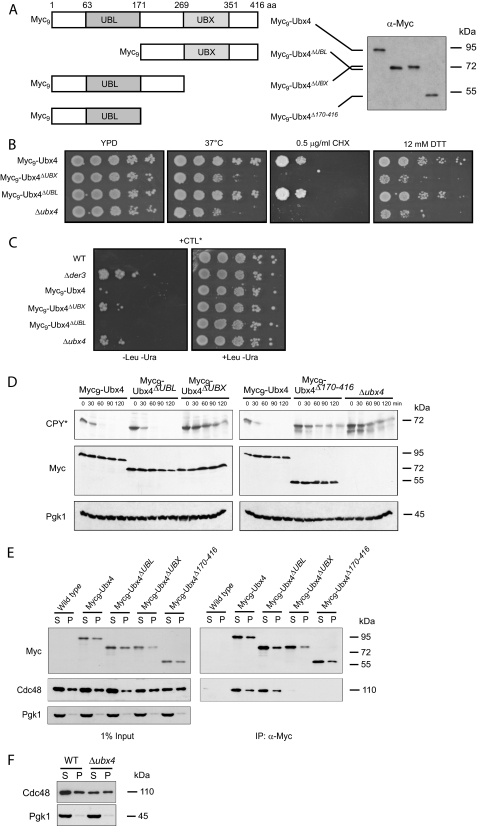
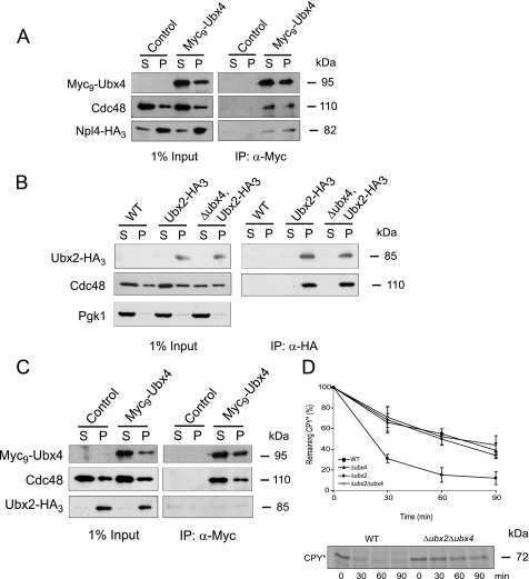
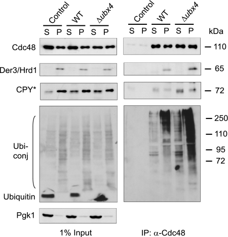
Misfolded proteins of the secretory pathway are recognized in the endoplasmic reticulum (ER), retrotranslocated into the cytoplasm, and degraded by the ubiquitin-proteasome system. Right after retrotranslocation and polyubiquitination, they are extracted from the cytosolic side of the ER membrane through a complex consisting of the AAA ATPase Cdc48 (p97 in mammals), Ufd1, and Npl4. This complex delivers misfolded proteins to the proteasome for final degradation. Extraction, delivery, and processing of ERAD (ER-associated degradation) substrates to the proteasome requires additional cofactors of Cdc48. Here we characterize the UBX domain containing protein Ubx4 (Cui1) as a crucial factor for the degradation of polyubiquitinated proteins via ERAD. Ubx4 modulates the Cdc48-Ufd1-Npl4 complex to guarantee its correct function. Mutant variants of Ubx4 lead to defective degradation of misfolded proteins and accumulation of polyubiquitinated proteins bound to Cdc48. We show the requirement of the UBX domain of Ubx4 for its function in ERAD. The observation that Ubx2 and Ubx4 are not found together in one complex with Cdc48 suggests several distinct steps in modulating the activity and localization of Cdc48 in ERAD.


Figures

FIGURE 1.
FIGURE 1.
Growth assays indicate a participation of Ubx4 in protein degradation. A, growth assays to analyze Δubx strains for sensitivity toward heat, cycloheximide (CHX), and dithiothreitol (DTT). 10-Fold serial dilutions of the indicated strains were spotted onto plates containing YPD, YPD plus cycloheximide, or YPD plus DTT and grown for 2–4 days at 30 °C or as indicated before being photographed. B, schematic representation of the ERAD substrate CTL*. C, growth test of yeast strains transformed with a plasmid expressing CTL* under the control of the GAL4 promotor. 10-Fold serial dilutions of the indicated strains were spotted onto plates containing selective media without or, as a control, with leucine and grown for 2–3 days at 30 °C before being photographed. The W303 prc1-1 wild type (WT) strain defective in the LEU2 gene fails to grow on medium lacking leucine, whereas strains defective in ERAD are able to grow due to complementation of the leucine deficiency by CTL*. The strain lacking the E3 ligase Der3/Hrd1 serves as a control. Two of the seven screened Δubx deletion strains, Δubx2 and Δubx4, complement the leucine auxotrophy.
FIGURE 2.
FIGURE 2.
Pulse-chase experiments and membrane extraction assays. A–C, pulse-chase analysis of CPY* and Ste6*-HA degradation. Wild type (WT) and Δubx4 (A and B) and wild type, Δubx2, Δubx4, and Δubx6 mutant cells (C) were grown and radiolabeled. Extracts were prepared, and the substrates were immunoprecipitated with specific antibodies at the indicated time points. Samples were separated via SDS-PAGE, and proteins were detected using a PhosphorImager system. D, cycloheximide chase experiment to analyze if Myc9-Ubx4 is functional regarding degradation of CPY* compared with wild type (WT). Pgk1 served as a loading control. Immunoblotting was done using specific antibodies. E, analysis of Ubx4 distribution in soluble and membrane fractions. Membrane extraction assay was performed in wild type cells expressing Myc9-Ubx4. Spheroblasted cells were homogenized and treated with different agents. Subsequently, the samples were separated by high speed centrifugation into supernatant (S) and pellet (P) fractions and analyzed via immunodetection with Myc, Cdc48, and as a control, Sec61 antibodies.
FIGURE 3.
FIGURE 3.
Function of the UBL and UBX domain of Ubx4 in ERAD. A, schematic representation of the Ubx4 domain structure and the deletions generated as well as their molecular masses determined by SDS-PAGE, subsequent Western blotting, and immunodetection using Myc antibodies. B, cells expressing Myc9-tagged Ubx4, Myc9-tagged fragments of Ubx4 deleted in the UBX or the UBL domain, as well as cells deleted in UBX4 were grown on different media as described in legend to Fig. 1A. C, growth test of cells expressing Myc9-tagged Ubx4, Myc9-tagged fragments of Ubx4 deleted in the UBX or the UBL domain, as well as cells deleted in UBX4, and for control, wild type and DER3 deleted cells were transformed with a plasmid expressing CTL* under control of the GAL4 promotor. The test was done as described in legend to Fig. 1C. D, cycloheximide chase experiments of CPY* degradation in different UBX4 deletion mutants. Detection of Myc signals served as control for expression and stability of Ubx4 and its fragments. Pgk1 (3-phosphoglycerate kinase) protein was used as a loading control. Immunoblotting was done using the respective specific antibodies. E, interaction of N-terminal Myc9-tagged Ubx4 and its truncated variants carrying deletions of the UBX or the UBL domain with Cdc48. Cells expressing Myc9-Ubx4, its Myc9-tagged truncated variants, and as a control wild type Ubx4 were lysed and separated into soluble (S) and membrane (P) proteins. Membrane proteins (P) were solubilized by digitonin treatment. Interaction of the Myc9-Ubx4 and mutant proteins with Cdc48 was followed by immunoprecipitation (IP) using Myc-specific antibodies. After SDS-PAGE and Western blotting, proteins were detected using specific antibodies. Pgk1 was used as a loading control. F, amount of Cdc48 in wild type (WT) cells compared with Δubx4 cells after fractionation of cell lysates by high speed centrifugation. Loading control was done using Pgk1 antibody.
FIGURE 4.
FIGURE 4.
Binding of cofactors to the Cdc48 complex. A, interaction of the Cdc48-Ubx4 complex with Npl4-HA3. Cells expressing Myc9-Ubx4 and Npl4-HA3 and, as a control, cells only expressing Npl4-HA3 were lysed and separated into soluble (S) and membrane (P) proteins. Membrane proteins (P) were solubilized by digitonin treatment. Immunoprecipitation (IP) of Myc9-Ubx4 (right panel) was done using monoclonal Myc antibody. Npl4-HA3 was detected using monoclonal HA antibody. 1% yeast extract (left panel) was immunoblotted for control. B, Cdc48-Ubx2 interaction is not affected in a Δubx4 strain. Ubx2-HA3 was expressed in wild type and Δubx4 cells and coprecipitated Cdc48. In the control strain Ubx2-HA3 was not expressed. Cell lysis and preparation of soluble and membrane proteins were done as described in A. Pgk1 was used as loading control. C, binding of Ubx4 excludes Ubx2 from the Cdc48 complex. Cells expressing Myc9-Ubx4 and Ubx2-HA3 and, as a control, cells only expressing Ubx2-HA3 were lysed and separated into soluble (S) and membrane (P) proteins. Cell lysis and preparation of soluble and membrane proteins were done as in A. Coimmunoprecipitation experiments were done using monoclonal Myc antibodies. Proteins were separated on SDS-PAGE and detected on immunoblots using Myc, Cdc48, or HA antibodies. D, pulse-chase analysis of CPY* degradation in wild type (WT), Δubx2, Δubx4, and Δubx2Δubx4 mutants. Cells were grown and radiolabeled; extracts were prepared, and the substrate was immunoprecipitated with specific CPY antibodies at the indicated time points. Samples were separated via SDS-PAGE, and the protein was detected using a PhosphorImager system.
FIGURE 5.
FIGURE 5.
Δubx4 mutants exhibit an increased load of CPY * and polyubiquitinated proteins on the Cdc48 complex. Wild type and Δubx4 cells were lysed and separated into soluble (S) and membrane (P) proteins. Membrane proteins (P) were solubilized by digitonin treatment. Coimmunoprecipitation (IP) of CPY*, Der3/Hrd1, and polyubiquitinated proteins with Cdc48 was done using Cdc48-specific antibodies. After SDS-PAGE and blotting onto nitrocellulose, protein detection was done using specific antibodies against CPY, Der3/Hrd1, and ubiquitin following ECL detection. Pgk1 was used as loading control.

References

    1. Hiller M. M., Finger A., Schweiger M., Wolf D. H. ( 1996) Science 273, 1725– 1728 - PubMed
    1. Kostova Z., Wolf D. H. ( 2003) EMBO J. 22, 2309– 2317 - PMC - PubMed
    1. Ahner A., Brodsky J. L. ( 2004) Trends Cell Biol. 14, 474– 478 - PubMed
    1. Schäfer A., Kostova Z., Wolf D. H. ( 2008) in The Ubiquitin-Proteasome System and Disease, ( Mayer R. J., Ciechanover A., Rechsteiner M. eds) pp. 123– 143, Wiley-VCH, Weinheim, Germany
    1. Raasi S., Wolf D. H. ( 2007) Semin. Cell Dev. Biol. 18, 780– 791 - PubMed

Publication types

MeSH terms

Substances