. 2009 May 29;284(22):15026-37.
doi: 10.1074/jbc.M109.004812. Epub 2009 Apr 9.

A methyltransferase-independent function for Rmt3 in ribosomal subunit homeostasis

Affiliations

A methyltransferase-independent function for Rmt3 in ribosomal subunit homeostasis

Audrey Perreault et al. J Biol Chem. .

Abstract

Schizosaccharomyces pombe Rmt3 is a member of the protein-arginine methyltransferase (PRMT) family and is the homolog of human PRMT3. We previously characterized Rmt3 as a ribosomal protein methyltransferase based on the identification of the 40 S Rps2 (ribosomal protein S2) as a substrate of Rmt3. RMT3-null cells produce nonmethylated Rps2 and show mis-regulation of the 40 S/60 S ribosomal subunit ratio due to a small subunit deficit. For this study, we have generated a series of RMT3 alleles that express various amino acid substitutions to characterize the functional domains of Rmt3 in Rps2 binding, Rps2 arginine methylation, and small ribosomal subunit production. Notably, catalytically inactive versions of Rmt3 restored the ribosomal subunit imbalance detected in RMT3-null cells. Consistent with a methyltransferase-independent function for Rmt3 in small ribosomal subunit production, the expression of an Rps2 variant in which the identified methylarginine residues were substituted with lysines showed normal levels of 40 S subunit. Importantly, substitutions within the zinc finger domain of Rmt3 that abolished Rps2 binding did not rescue the 40 S ribosomal subunit deficit of RMT3-null cells. Our findings suggest that the Rmt3-Rps2 interaction, rather than Rps2 methylation, is important for the function of Rmt3 in the regulation of small ribosomal subunit production.


Figures

FIGURE 1.
FIGURE 1.
Schematic representation of S. pombe Rmt3 and substitutions introduced in this study. The predicted C2H2 zinc finger motif, conserved regions CR1 and CR2, protein methyltransferase (MTase) motifs (Post-I, II, and III), and the THW loop region of Rmt3 are shown. Asterisks are present below the canonical cysteine and histidine residues of the zinc finger motif. The arrows indicate the positions of the various substitutions and amino acid deletion introduced to fission yeast Rmt3. Amino acid numbering is from S. pombe Rmt3. Sequence alignments are from Neurospora crassa (Nc), Aspergillus nidulans (An), S. pombe (Sp), Cryptococcus neoformans (Cn), Homo sapiens (Hs), and Drosophila melanogaster (Dm).
FIGURE 2.
FIGURE 2.
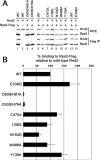
The integrity of the Rmt3 zinc finger motif is essential for the association with Rps2. A, immunoblot analysis of whole cell extracts (WCE) and FLAG immunoprecipitates (IP) prepared from RMT3-null cells (lane 6) that expressed various alleles of RMT3 (lanes 1–5 and 7–13) and that were previously transformed with a plasmid expressing Rps2-FLAG (lanes 1 and 3–13)or vector control (lane 2). Antibodies for Western blotting were rabbit polyclonal and mouse monoclonal antibodies specific to Rmt3 and the FLAG epitope, respectively. B, quantification of at least three independent copurification experiments as shown in A. WT, wild type.
FIGURE 3.
FIGURE 3.
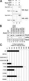
Glutamic acid 338 within motif II of the methyltansferase domain and tryptophan 488 of the THW loop are required for the catalytic activity of Rmt3. A, a methylarginine-specific antibody that specifically recognizes methylated Rps2. Shown is Western blot analysis of total cell lysates prepared from wild-type cells (lane 1), RMT3-null cells (lane 2), and RPS2-null cells that were previously transformed with a plasmid expressing Rps2-GFP (lane 3). Antibodies for Western blotting (WB) were Rps2-specific and methylarginine-specific (mRG) polyclonal antibodies (top and bottom, respectively). B, Western blot analysis of total cell lysates prepared from RMT3-null cells (lane 2) that expressed various alleles of RMT3 (lanes 1 and 3–10). Antibodies for Western blotting were Rmt3-specific, methylarginine-specific, and Rps2-specific rabbit polyclonal antibodies (top, middle, and bottom, respectively). C, percentage of Rps2 methylation as determined from at least three independent experiments as shown in B. WT, wild type.
FIGURE 4.
FIGURE 4.
Methyltransferase activity of the different Rmt3 variants assayed in vitro. An equal amount of immunopurified Rps2 (lanes 1–10) was incubated with GST alone (lane 1) or with GST fused to the different Rmt3 variants (lanes 2–10) in the presence of 1.1 μCi of 3H-labeled S-adenosyl-l-methionine for 3 h at 30 °C. Proteins were separated by SDS-PAGE and stained with Coomassie Blue (top). The stained gel was dried and subjected to fluorography (bottom). The position of recombinant GST-Rmt3 proteins and methylated Rps2 is shown on the right of the upper panel and lower panel, respectively. WT, wild type.
FIGURE 5.
FIGURE 5.
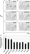
Catalytically inactive alleles of RMT3 restore the ribosomal subunit imbalance detected in RMT3-null cells. Shown is sucrose gradient analysis of total extracts prepared from RMT3-null cells (A) that expressed wild-type (B), C63S/H76A (C), C60S/H81A (D), E338Q (E), and W488A (F) alleles of RMT3. G, quantification of the 60 S/40 S free ribosomal subunit ratio for the different RMT3 alleles as determined from at least three independent experiments. Asterisks above bars indicate statistically significant increase relative to wild-type Rmt3 (p < 0.0001), as determined by Student's t tests.
FIGURE 6.
FIGURE 6.
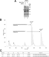
Identification of methylarginine residues of fission yeast Rps2 by MS. A, large scale purification of fission yeast Rps2. Coomassie Blue staining of FLAG immunoprecipitates prepared from extracts of strain FBY154 (lane 1) and FBY157 (lane 2). The arrowheads indicate the position of Rps2-FLAG. Molecular mass markers are indicated on the right in kDa. B, MS spectrum of two asymmetrically dimethylated peptides from S. pombe Rps2. Asterisks and diamonds mark those peaks in the MS spectrum that have been correlated with dimethylarginine modifications, as a result of signal generation in the ion precursor scan at m/z 46. The peptide sequence is shown beside each dimethylated peptide, as verified by accurate mass measurement. C, arginine-methylated residues found in S. pombe Rps2. The positions of the two different methylated peptides are in amino acids. MC, missed cleavages. IP, immunoprecipitation.
FIGURE 7.
FIGURE 7.
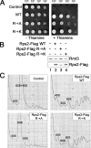
Arginine methylation is not required for the function of Rps2 in ribosome biogenesis. A, 10-fold serial dilutions of strains FBY136 (control), FBY217 (wild type; WT), FBY218 (Arg → Ala), and FBY219 (Arg → Lys) were spotted onto Edinburgh minimal medium plates with (right) or without (left) thiamine. B, Western blot analysis of total cell lysates prepared from strain FBY136 (lane 1), FBY217 (lane 2), FBY218 (lane 3), and FBY219 (lane 4). Antibodies for Western blotting were a rabbit polyclonal anti-Rmt3 (top) and a mouse monoclonal anti-FLAG (bottom). C, sucrose gradient analysis of total extracts prepared from strains FBY136 (control), FBY217 (wild type), FBY218 (Arg → Ala), and FBY219 (Arg → Lys).

References

    1. Bachand, F. (2007) Eukaryot. Cell 6 889-898 - PMC - PubMed
    1. Bedford, M. T., and Richard, S. (2005) Mol. Cell 18 263-272 - PubMed
    1. Bedford, M. T. (2007) J. Cell Sci. 120 4243-4246 - PubMed
    1. Gary, J. D., and Clarke, S. (1998) Prog. Nucleic Acid Res. Mol. Biol. 61 65-131 - PubMed
    1. Kwak, Y. T., Guo, J., Prajapati, S., Park, K. J., Surabhi, R. M., Miller, B., Gehrig, P., and Gaynor, R. B. (2003) Mol. Cell 11 1055-1066 - PubMed

Publication types

MeSH terms

LinkOut - more resources