

The effects of cycling levels of 17beta-estradiol and progesterone on the magnitude of temporomandibular joint-induced nociception
- PMID: 19359384
- PMCID: PMC2717863
- DOI: 10.1210/en.2008-1707
The effects of cycling levels of 17beta-estradiol and progesterone on the magnitude of temporomandibular joint-induced nociception
Abstract
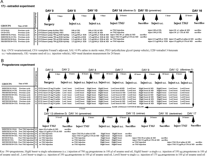
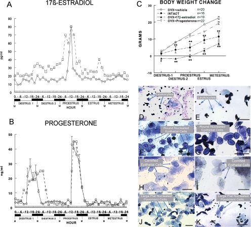
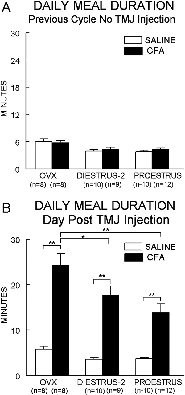
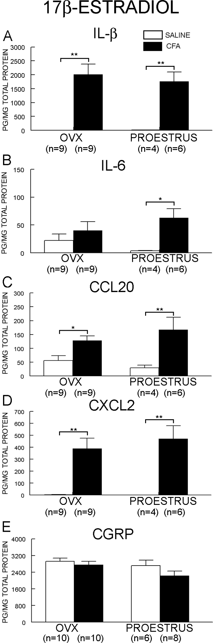
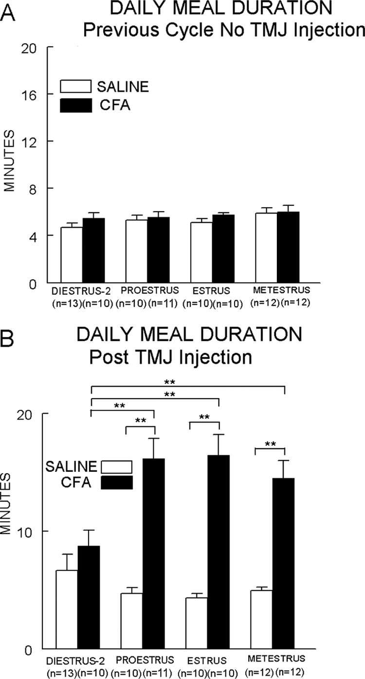
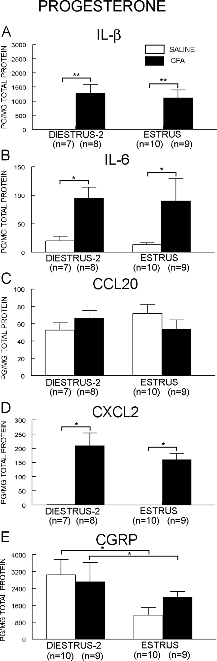
A greater incidence of temporomandibular joint (TMJ) pain is reported in females, suggesting that gonadal hormones may play a role in this condition. However, the exact roles of 17beta-estradiol (E2) and progesterone (P4) in TMJ pain are not completely known. Two experiments were performed to determine the separate roles of E2 and P4 in TMJ nociception at various stages of the estrous cycle. Ovariectomized (OVX) rats were cycled with physiological concentrations of E2 or P4. The E2-cycled rats then received bilateral TMJ injections of saline (SAL) or complete Freund's adjuvant (CFA) on the morning of diestrus-2 (low E2 condition) or proestrus (high E2 condition). As a control, OVX rats (no ovarian E2 and no replacement) were injected with SAL or CFA. The TMJ nociception was measured using a validated novel method in which an increase in meal duration directly correlated to the intensity of deep TMJ nociception. In the E2 experiment, CFA injection, but not SAL, increased TMJ nociception in the OVX group, but the effect was less pronounced in diestrus-2 and even less in proestrus. In the P4 experiment, the rats receiving TMJ CFA in diestrus-2 (end of minor P4 surge) did not show an increase in TMJ nociception, whereas the rats injected in proestrus (major P4 surge), estrus (low P4), and metestrus (low P4) had similar increases in TMJ nociception. The hormones' concentration did not affect TMJ IL-1beta, IL-6, C-C motif ligand 20, or C-X-C motif ligand 2 or the trigeminal ganglia calcitonin gene-related peptide. The high physiological concentrations of E2 observed at proestrus and the low P4 concentrations observed at diestrus-2 attenuated or eliminated CFA-induced TMJ nociception. The results suggest that the cyclic estrous cycle concentrations of E2 and P4 can influence CFA-induced TMJ nociception in the rat.
Figures
Similar articles
-
Modulation of temporomandibular joint nociception and inflammation in male rats after administering a physiological concentration of 17β-oestradiol.Eur J Pain. 2013 Feb;17(2):174-84. doi: 10.1002/j.1532-2149.2012.00183.x. Epub 2012 Jun 19. Eur J Pain. 2013. PMID: 22715057 Free PMC article.
-
The influence of sex and ovarian hormones on temporomandibular joint nociception in rats.J Pain. 2008 Jul;9(7):630-8. doi: 10.1016/j.jpain.2008.02.006. J Pain. 2008. PMID: 18420460
-
Estradiol upregulates voltage-gated sodium channel 1.7 in trigeminal ganglion contributing to hyperalgesia of inflamed TMJ.PLoS One. 2017 Jun 5;12(6):e0178589. doi: 10.1371/journal.pone.0178589. eCollection 2017. PLoS One. 2017. PMID: 28582470 Free PMC article.
-
Regulation of object recognition and object placement by ovarian sex steroid hormones.Behav Brain Res. 2015 May 15;285:140-57. doi: 10.1016/j.bbr.2014.08.001. Epub 2014 Aug 15. Behav Brain Res. 2015. PMID: 25131507 Free PMC article. Review.
-
"Testosterone decreases temporomandibular joint nociception"- A systematic review of studies on animal models.Arch Oral Biol. 2022 Jul;139:105430. doi: 10.1016/j.archoralbio.2022.105430. Epub 2022 Apr 14. Arch Oral Biol. 2022. PMID: 35461068
Cited by
-
The degeneration-pain relationship in the temporomandibular joint: Current understandings and rodent models.Front Pain Res (Lausanne). 2023 Feb 9;4:1038808. doi: 10.3389/fpain.2023.1038808. eCollection 2023. Front Pain Res (Lausanne). 2023. PMID: 36846071 Free PMC article. Review.
-
Activation of estrogen receptor α enhances bradykinin signaling in peripheral sensory neurons of female rats.J Pharmacol Exp Ther. 2014 Jun;349(3):526-32. doi: 10.1124/jpet.114.212977. Epub 2014 Apr 4. J Pharmacol Exp Ther. 2014. PMID: 24706985 Free PMC article.
-
Estrogen in cycling rats alters gene expression in the temporomandibular joint, trigeminal ganglia and trigeminal subnucleus caudalis/upper cervical cord junction.J Cell Physiol. 2011 Dec;226(12):3169-80. doi: 10.1002/jcp.22671. J Cell Physiol. 2011. PMID: 21321935 Free PMC article.
-
Estrogen receptor β activation is antinociceptive in a model of visceral pain in the rat.J Pain. 2012 Jul;13(7):685-94. doi: 10.1016/j.jpain.2012.04.010. Epub 2012 Jun 13. J Pain. 2012. PMID: 22698981 Free PMC article.
-
Knockdown of Fcγ receptor III in an arthritic temporomandibular joint reduces the nociceptive response in rats.Arthritis Rheum. 2010 Oct;62(10):3109-18. doi: 10.1002/art.27630. Arthritis Rheum. 2010. PMID: 20589683 Free PMC article.
References
-
- Lipton JA, Ship JA, Larach-Robinson D 1993 Estimated prevalence and distribution of reported orofacial pain in the United States. J Am Dent Assoc 124:115–121 - PubMed
-
- LeResche L 1997 Epidemiology of temporomandibular disorders: implications for the investigation of etiologic factors. Crit Rev Oral Biol Med 8:291–305 - PubMed
-
- Fillingim RB, Ness TJ 2000 Sex-related hormonal influences on pain and analgesic responses. Neurosci Biobehav Rev 24:485–501 - PubMed
-
- Shinal RM, Fillingim RB 2007 Overview of orofacial pain: epidemiology and gender differences in orofacial pain. Dent Clin North Am 51:1–18, v - PubMed
-
- Goulet JP, Lavigne GJ, Lund JP 1995 Jaw pain prevalence among French-speaking Canadians in Quebec and related symptoms of temporomandibular disorders. J Dent Res 74:1738–1744 - PubMed
Publication types
MeSH terms
Substances
Grants and funding
LinkOut - more resources
Full Text Sources
Medical
Research Materials
