. 2009 Jun;296(6):G1167-79.
doi: 10.1152/ajpgi.90496.2008. Epub 2009 Apr 9.

A novel Toll-like receptor 4 antagonist antibody ameliorates inflammation but impairs mucosal healing in murine colitis

Affiliations

A novel Toll-like receptor 4 antagonist antibody ameliorates inflammation but impairs mucosal healing in murine colitis

Ryan Ungaro et al. Am J Physiol Gastrointest Liver Physiol. 2009 Jun.

Abstract

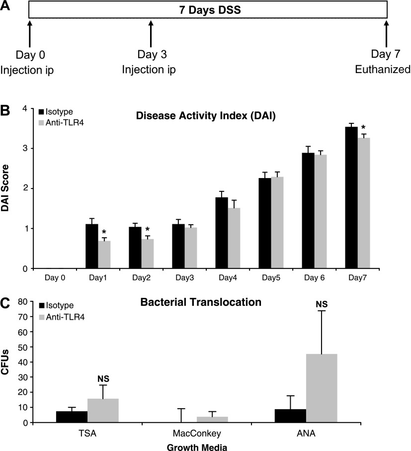
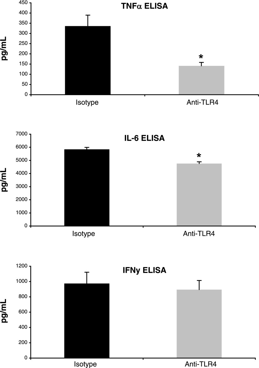
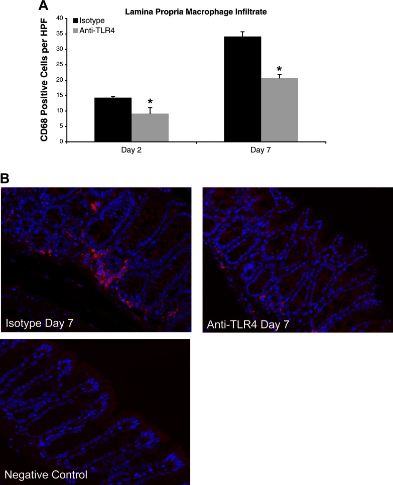
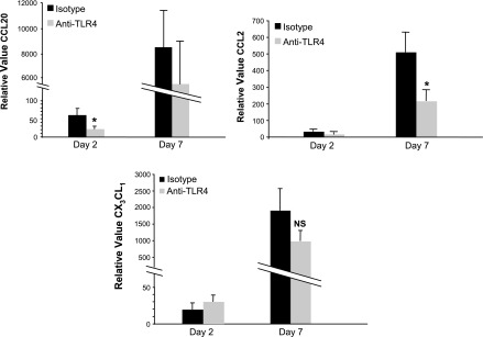
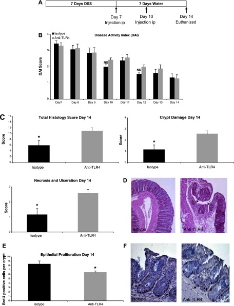
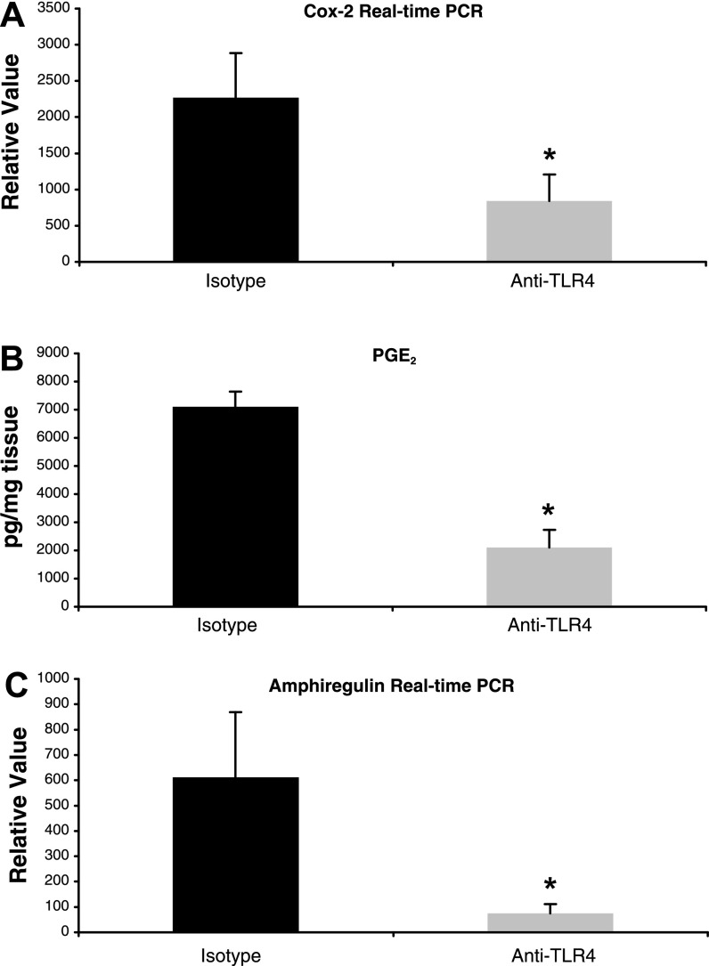
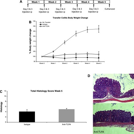
Dysregulated innate immune responses to commensal bacteria contribute to the development of inflammatory bowel disease (IBD). TLR4 is overexpressed in the intestinal mucosa of IBD patients and may contribute to uncontrolled inflammation. However, TLR4 is also an important mediator of intestinal repair. The aim of this study is to examine the effect of a TLR4 antagonist on inflammation and intestinal repair in two murine models of IBD. Colitis was induced in C57BL/6J mice with dextran sodium sulfate (DSS) or by transferring CD45Rb(hi) T cells into RAG1-/- mice. An antibody (Ab) against the TLR4/MD-2 complex or isotype control Ab was administered intraperitoneally during DSS treatment, recovery from DSS colitis, or induction of colitis in RAG1-/- mice. Colitis severity was assessed by disease activity index (DAI) and histology. The effect of the Ab on the inflammatory infiltrate was determined by cell isolation and immunohistochemistry. Mucosal expression of inflammatory mediators was analyzed by real-time PCR and ELISA. Blocking TLR4 at the beginning of DSS administration delayed the development of colitis with significantly lower DAI scores. Anti-TLR4 Ab treatment decreased macrophage and dendritic cell infiltrate and reduced mucosal expression of CCL2, CCL20, TNF-alpha, and IL-6. Anti-TLR4 Ab treatment during recovery from DSS colitis resulted in defective mucosal healing with lower expression of COX-2, PGE(2), and amphiregulin. In contrast, TLR4 blockade had minimal efficacy in ameliorating inflammation in the adoptive transfer model of chronic colitis. Our findings suggest that anti-TLR4 therapy may decrease inflammation in IBD but may also interfere with colonic mucosal healing.


Figures

Fig. 1.
Fig. 1.
TLR4 blockade ameliorates DSS-induced colitis. A: treatment schedule for anti-TLR4 antibody and isotype control during 2.5% DSS administration. Mice were injected intraperitoneally (ip) at the indicated time points. B: mice treated with anti-TLR4 antibody had reduced severity of colitis as measured by disease activity index (DAI). Data represent means ± SE of 3 independent experiments with a total of 24 mice (n = 15 for anti-TLR4 and n = 9 for isotype control-treated mice). *P < 0.05. C: bacterial translocation as measured by culture of mesenteric lymph nodes (MLNs) on indicated media from anti-TLR4-treated (n = 4) and isotype-treated (n = 4) mice. Data represent means ± SE of 2 independent experiments. CFUs, colony-forming units; TSA, Trypticase soy agar; ANA, anaerobic with PEA; NS, not significant.
Fig. 2.
Fig. 2.
Inhibition of TLR4 decreases production of proinflammatory cytokines. Mucosal production of cytokines at day 7 of DSS treatment was measured by ELISA using colon culture supernatants. 100 mg of tissue fragments from each part of the colon were cultured for 24 h in 1 ml of serum-free RPMI 1640 from anti-TLR4-treated (n = 9) and isotype-treated (n = 8) mice. Data represent mean values ± SE of supernatants measured in duplicate wells from 3 independent experiments. *P < 0.05.
Fig. 3.
Fig. 3.
Anti-TLR4 antibody treatment reduces lamina propria macrophage infiltrate. A: number of lamina propria macrophages per high-power field (HPF) at different time points of DSS administration. CD68 staining in anti-TLR4-treated (day 2: n = 3; day 7: n = 9) and isotype-treated (day 2: n = 3; day 7: n = 8) mice was evaluated in at least 5 different areas per mouse at ×400 magnification. Data are expressed as mean values ± SE from 3 independent experiments. Day 2 and day 7 correspond to the treatment schedule in Fig. 1A. *P < 0.05. B: representative photos of immunofluorescent staining of CD68-positive cells at ×400 magnification on day 7 of DSS treatment. The primary antibody was omitted for negative control slides.
Fig. 4.
Fig. 4.
TLR4 signaling contributes to expression of chemokines involved in the recruitment of antigen presenting cells (APCs). Real-time PCR results for mucosal expression of CCL20, CCL2, and CX3CL1 in anti-TLR4 antibody-treated (day 2: n = 3; day 7: n = 9) and isotype control-treated (day 2: n = 3; day 7: n = 8) mice. Data are presented as mean ± SE relative values of expression from 2 independent experiments with duplicate samples. Day 2 and day 7 correspond to the treatment schedule in Fig. 1A. *P < 0.05.
Fig. 5.
Fig. 5.
TLR4 blockade during recovery from DSS colitis impairs mucosal healing and epithelial proliferation. A: treatment schedule for anti-TLR4 antibody and isotype control during recovery from DSS colitis. Mice were injected intraperitoneally (ip) at the indicated time points. B: there were no clinical differences between mice treated with anti-TLR4 antibody or isotype control during recovery from DSS colitis as measured by DAI. Data represent the mean ± SE of 2 independent experiments with a total of 22 mice (n = 12 for anti-TLR4 and n = 10 for isotype control-treated mice). C: histological assessment of colitis severity in anti-TLR4 antibody- and isotype control-treated mice. Data represent the mean histological score ± SE for mice from 2 independent experiments (n = 4 for anti-TLR4 and n = 4 for isotype). Anti-TLR4 antibody-treated mice failed to recover from DSS-induced damage with higher total histological damage scores at day 14. Specifically, whereas isotype control mice had begun to repair mucosal damage caused by DSS, the anti-TLR4 antibody-treated mice had persistent crypt damage and ulceration at day 14. *P < 0.05. D: representative microscopic pictures of hematoxylin and eosin (H&E) staining of colon sections from anti-TLR4 antibody and isotype control-treated mice. Arrows indicate areas of persistent crypt damage and ulceration in an anti-TLR4-treated mouse colon section. E: epithelial proliferation was measured by bromodeoxyuridine (BrdU) labeling index (number of BrdU-positive cells per crypt at ×400 magnification). Data represent means ± SE for mice from 2 independent experiments (n = 4 for anti-TLR4 and n = 4 for isotype). *P < 0.05. F: representative photo of immunohistochemical staining of incorporated BrdU into colonic epithelium. Proliferating cells labeled with BrdU have brown nuclei as indicated by the arrows.
Fig. 6.
Fig. 6.
Anti-TLR4 antibody treatment results in decreased expression of mediators of mucosal repair. A: real-time PCR results of mucosal expression of COX-2 in anti-TLR4 antibody-treated (n = 4) and isotype control-treated (n = 4) mice at day 14. Data are presented as mean ± SE relative values of expression from 2 independent experiments with duplicate samples. *P < 0.05. B: ELISA results for PGE2 levels in colon culture supernatants from anti-TLR4 antibody-treated (n = 4) and isotype control-treated (n = 4) mice at day 14. Data represent mean values ± SE of supernatants measured in triplicate from 2 independent experiments. *P < 0.05. C: real-time PCR results of mucosal expression of amphiregulin in anti-TLR4 antibody-treated (n = 4) and isotype control-treated (n = 4) mice at day 14. Data are presented as mean ± SE relative values of expression from 2 independent experiments with duplicate samples. *P < 0.05.
Fig. 7.
Fig. 7.
Anti-TLR4 antibody treatment has minimal effect in the transfer model of chronic colitis. A: treatment schedule for anti-TLR4 antibody and isotype control during adoptive transfer model of colitis. CD4+CD62L+ T cells were injected ip into RAG1−/− mice at day 0 of week 1. Mice were then injected ip at the indicated time points with either anti-TLR4 antibody or isotype control and euthanized at the end of 5 wk. B: anti-TLR4 treatment does not result in significant clinical differences. Graph shows percent body weight change. Data represent the average ± SE of 2 independent experiments with a total of 18 mice (n = 9 for anti-TLR4 and n = 9 for isotype-treated mice). Top line represents normal growth of control RAG−/− mice that did not undergo T cell transfer (n = 4). C: histological assessment of colitis severity in anti-TLR4 antibody and isotype control-treated mice. Data represent the mean histological score ± SE for mice from 2 independent experiments (n = 4 for anti-TLR4 and n = 4 for isotype). Anti-TLR4-treated mice have slightly worse colitis on histology. D: representative microscopic pictures of H&E staining of colon sections from anti-TLR4 antibody and isotype control-treated mice.

References

    1. Allison MC, Poulter LW. Changes in phenotypically distinct mucosal macrophage populations may be a prerequisite for the development of inflammatory bowel disease. Clin Exp Immunol 85: 504–509, 1991. - PMC - PubMed
    1. Ancuta P, Rao R, Moses A, Mehle A, Shaw SK, Luscinskas FW, Gabuzda D. Fractalkine preferentially mediates arrest and migration of CD16+ monocytes. J Exp Med 197: 1701–1707, 2003. - PMC - PubMed
    1. Aranda R, Sydora BC, McAllister PL, Binder SW, Yang HY, Targan SR, Kronenberg M. Analysis of intestinal lymphocytes in mouse colitis mediated by transfer of CD4+, CD45RBhigh T cells to SCID recipients. J Immunol 158: 3464–3473, 1997. - PubMed
    1. Baumgart DC, Metzke D, Schmitz J, Scheffold A, Sturm A, Wiedenmann B, Dignass AU. Patients with active inflammatory bowel disease lack immature peripheral blood plasmacytoid and myeloid dendritic cells. Gut 54: 228–236, 2005. - PMC - PubMed
    1. Bennett-Guerrero E, Grocott HP, Levy JH, Stierer KA, Hogue CW, Cheung AT, Newman MF, Carter AA, Rossignol DP, Collard CD. A phase II, double-blind, placebo-controlled, ascending-dose study of Eritoran (E5564), a lipid A antagonist, in patients undergoing cardiac surgery with cardiopulmonary bypass. Anesth Analg 104: 378–383, 2007. - PubMed

Publication types

MeSH terms

LinkOut - more resources