. 2009 Apr 28;106(17):7119-24.
doi: 10.1073/pnas.0902745106. Epub 2009 Apr 9.

IL-1 acts directly on CD4 T cells to enhance their antigen-driven expansion and differentiation

Affiliations

IL-1 acts directly on CD4 T cells to enhance their antigen-driven expansion and differentiation

Shlomo Z Ben-Sasson et al. Proc Natl Acad Sci U S A. .

Abstract

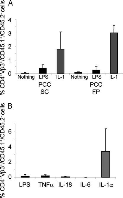
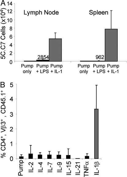
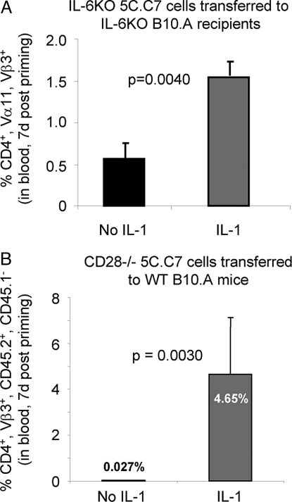
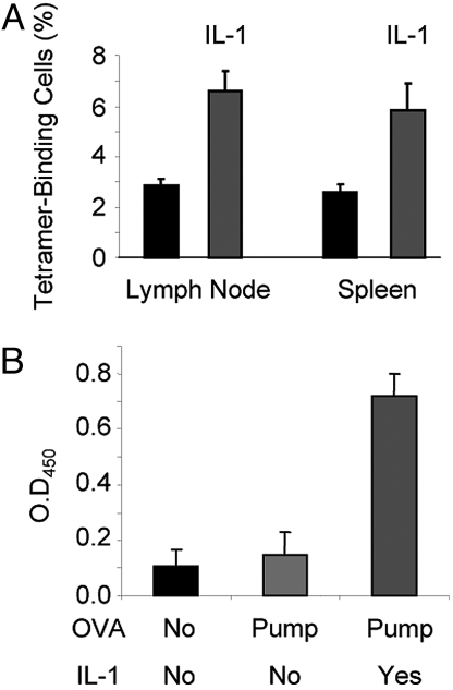
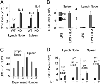
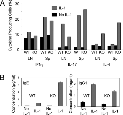
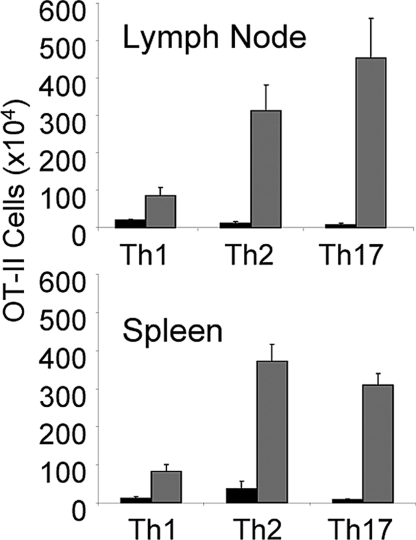
IL-1 causes a marked increase in the degree of expansion of naïve and memory CD4 T cells in response to challenge with their cognate antigen. The response occurs when only specific CD4 T cells can respond to IL-1beta, is not induced by a series of other cytokines and does not depend on IL-6 or CD-28. When WT cells are primed in IL-1R1(-/-) recipients, IL-1 increases the proportion of cytokine-producing transgenic CD4 T cells, especially IL-17- and IL-4-producing cells, strikingly increases serum IgE levels and serum IgG1 levels. IL-1beta enhances antigen-mediated expansion of in vitro primed Th1, Th2, and Th17 cells transferred to IL-1R1(-/-) recipients. The IL-1 receptor antagonist diminished responses to antigen plus lipopolysaccharide (LPS) by approximately 55%. These results indicate that IL-1beta signaling in T cells markedly induces robust and durable primary and secondary CD4 responses.


Conflict of interest statement

The authors declare no conflict of interest.

Figures

Fig. 1.
Fig. 1.
IL-1 augments primary responses. (A) IL-1 is superior to LPS in augmenting primary immune responses in vivo. The 5C.C7/RAG-2−/−/CD45.1 B10.A lymph node cells (1.5 × 10⁶) were transferred IP into CD45.2 B10.A mice. Ten days later, mice were immunized by s.c. or footpad (FP) injection of 500 μg of PCC only, with 25 μg of LPS or with rIL-1β (10 μg) in a miniosmotic pump. One week after priming, the percentage of CD4+/Vβ3+/CD45.1+/CD45.2 (i.e., 5C.C7) cells (Mean + SEM) among the PBMCs was determined as described in Materials and Methods. (B) IL-1 is superior to LPS and to other proinflammatory cytokines in augmenting primary immune responses in vivo. The 5C.C7/RAG-2−/−/CD45.1 B10.A lymph node (1 × 10⁶) and spleen (2.5 × 10⁶) cells were transferred IP into CD45.2 B10.A mice. Seven days later, mice were immunized by implantation of miniosmotic pumps containing 1 mg of PCC with either rIL-1α (10 μg), rIL-6 (10 μg), rIL-18 (10 μg), rTNFα (10 μg) or LPS (25 μg). One week after priming, the percentage of CD4+/Vβ3+/CD45.1+/CD45.2 cells among the PBMCs was determined as described in Materials and Methods.
Fig. 2.
Fig. 2.
The IL-1 effect persists and IL-1 acts on memory cells. (A) 5C.C7 cells primed in the presence of IL-1 persist. The 5C.C7/RAG-2−/−/CD45.2 B10.A lymph node (1 × 10⁶) and spleen (2 × 10⁶) cells were transferred IP into CD45.1 B10.A mice. Seven days later, mice were immunized by implantation of miniosmotic pumps containing PCC (1 mg) alone or with LPS (25 μg) or IL-1 (10 μg). Sixteen months later, the number of CD4+/Vβ3+/CD45.2+ cells in lymph nodes and spleens was determined. (B) IL-1, but not other cytokines, induces robust secondary responses in vivo. The 5C.C7/RAG-2−/−/CD45.1 B10.A lymph node (1 × 10⁶) and spleen (2 × 10⁶) cells were transferred IP into CD45.2 B10.A mice. Seven days later, mice were immunized by s.c. injection of 1 mg of PCC with 25 μg of LPS. Seven months after priming, the mice were boosted by implantation of miniosmotic pumps containing 1 mg of PCC with either rIL-1β (10 μg), rIL-2 (10 μg), rIL-4 (10 μg), rIL-7 (10 μg), rIL-9 (10 μg), rIL-15 (10 μg), rIL-21 (10 μg), rTNFα (10 μg), or no additional cytokine. One week after the boost, the percentage of CD4+/Vβ3+/CD45.1+ cells among PBMCs was determined.
Fig. 3.
Fig. 3.
Neither IL-6 or CD28 are needed. (A) IL-6 is not essential for the IL-1-mediated enhancement of primary immune responses. The 5C.C7/RAG-2−/−/IL-6−/− B10.A lymph node cells (1.5 × 10⁵) were transferred IP into CD3ε−/−, IL-6−/− B10.A mice. Eight days later, mice were immunized by implantation of miniosmotic pumps containing 1 mg of PCC with or without rIL-1β (10 μg). One week after priming, the percentage of CD4+/Vα11+/Vβ3+ cells among PBMCs was determined as described in Materials and Methods. (B) IL-1 enhances the response of antigen-stimulated CD28−/− 5C.C7 cells. The 5C.C7/RAG-2−/−/CD28−/− CD45.2 B10.A lymph node cells (1 × 10⁶) were transferred IP into normal CD45.1 B10.A mice. Eight days later, mice were immunized by implantation of miniosmotic pumps containing 500 μg of PCC with or without rIL-1β (10 μg). After 7 days, the percentage of CD4+/Vβ3+/CD45.2+/CD45.1 cells among PBMCs was determined as described in Materials and Methods.
Fig. 4.
Fig. 4.
IL-1 enhances polyclonal responses. (A) IL-1 increases the frequency of tetramer-binding CD4 cells in mice immunized with an LCMV peptide. C57BL/6 mice were immunized by implantation of miniosmotic pumps containing 100 μg of the gp61–80 LCMV peptide with or without rIL-1β(10 μg). After 7 days, the percentage of cells stained with the gp66–77 tetramer among the CD4+/Vα2+/CD44high population in the lymph nodes and spleens was determined. (B) IL-1 enhances the IgG1 anti-ovalbumin antibody response. C57BL/6 mice were immunized by implantation of miniosmotic pumps containing 1 mg ovalbumin with or without rIL-1β(10 μg). Sera were obtained 14 days after immunization and IgG1 anti-ovalbumin antibodies were determined at a 1:100 dilution by sandwich ELISA.
Fig. 5.
Fig. 5.
IL-1 acts on T cells. (A) IL-1 enhances responses of WT T cells in IL-1R1−/− recipients. OT-II/RAG1−/− C57BL/6 lymph node cells (5 × 10⁵) enriched for CD4 T cells, were transferred IP into WT or IL-1R1−/− (KO) C57BL/6 mice. Seven days later, mice were immunized by injection of 1 mg of OVA s.c. with LPS (25 μg) with or without rIL-1β (10 μg) in a miniosmotic pump. Seven days after immunization, the number and percentage of CD4+/Vα2+/Vβ5+ cells in the lymph nodes and spleens were determined as described in Materials and Methods. (B) IL-1-induced expansion of antigen-stimulated CD4 cells is cell autonomous. OT-II/RAG1−/− CD45.1 C57BL/6 and IL-1R1−/− OT-II/RAG1−/− C57BL/6 lymph node cells were transferred IP into B6 1L-1R1−/− Rag2−/− mice. Nine days later, mice were immunized by injection of 500 μg of OVA and 25 μg of LPS s.c. with or without rIL-1β (5 μg) administered through a miniosmotic pump. Seven days after immunization, the percentage of CD45.1 and CD45.2 CD4+/Vα2+/Vβ5+ cells in the lymph nodes and spleens was determined as described in Materials and Methods and the number of WT and IL-1R1−/− (KO) OT-II cells in these organs calculated. (C) IL-1 enhances responses of WT T cells in 1L-1R1−/− Rag1−/− recipients. OT-II/RAG1−/− C57BL/6 lymph node cells (5 × 10⁵) that had been enriched for CD4 T cells were transferred IP into B6 1L-1R1−/− Rag1−/− mice. Seven to 9 days later, mice were immunized by injection of 500 μg of OVA s.c. with LPS (25 μg) with or without rIL-1β (5 μg) that was administered in a miniosmotic pump. Seven days after immunization, the number and percentage of CD4+/Vα2+/Vβ5+ cells in the lymph nodes and spleens were determined as described in Materials and Methods. The ratios of OT-II cells in mice immunized with and without IL-1 from 3 independent experiments are presented. (D) IL-1 induces less expansion of antigen-stimulated IL-1R1−/− OT-II cells than of WT OT-II cells in WT recipients. OT-II/RAG1−/− C57BL/6 (WT) or IL-1R1−/− OT-II/RAG1−/− C57BL/6 (KO) lymph node cells were transferred IP into WT C57BL/6 mice. Nine days later, mice were immunized by injection of 500 μg of OVA and 25 μg of LPS s.c. with or without rIL-1β (5 μg) in a miniosmotic pump. Seven days after immunization, the percentage of CD4+/Vα2+/Vβ5+ cells in the lymph nodes and spleens was determined as described in Materials and Methods, and the number of OT-II cells the organs was calculated.
Fig. 6.
Fig. 6.
IL-1 enhances cytokine, IgE and IgG1 responses. (A) IL-1 augments the differentiation of cytokine-producing cells in OT-II cells primed in IL-1R1−/− recipients. Lymph nodes and spleens from mice of the experiment described in the legend to Fig. 5A were removed 7 days after immunization and the single cell suspensions were stimulated with PMA and ionomycin as described in Materials and Methods. The percentage of IL-4, IFNγ and IL-17-producing cells among the OT-II cells was determined by intracellular staining and FACS analysis of the CD4+, Vα2+, Vβ5+ gated population. (B) IL-1 induces the production of high levels of IgE and IgG1 7 days after priming in IL-1R1−/− mice. Mice from the experiment described in the legend to Fig. 5A were bled 7 days after immunization and the concentrations of serum IgG1, and IgE measured by ELISA.
Fig. 7.
Fig. 7.
IL-1 enhances the antigen-induced expansion of Th1, Th2, and Th17 cells in vivo. In vitro differentiated Th1, Th2, or Th17 OT-II cells were transferred IP into B6 1L-1R1−/− Rag1−/− mice (0.8 to 2 × 10⁶ per mouse). Seven days later, mice were immunized by implantation of a miniosmotic pump containing 1 mg of ovalbumin and either IL-1β (10 μg) or PBS. After a week, the number of CD4+/Vα2+, Vβ5+ cells in the lymph nodes and spleens was determined as described in Materials and Methods.

References

    1. Joffre O, Nolte MA, Spörri R, Reis e Sousa C. Inflammatory signals in dendritic cell activation and the induction of adaptive immunity. Immunol Rev. 2009;227:234–247. - PubMed
    1. Dinarello CA. Interleukin-1 family of ligands and receptors. In: Paul WE, editor. Fundamental Immunology. 6th Ed. Philadelphia: Lippincott Williams & Wilkins;
    1. Chen Q, Sen G, Snapper CM. Endogenous IL-1R1 signaling is critical for cognate CD4+ T cell help for induction of in vivo type 1 and type 2 antipolysaccharide and antiprotein Ig isotype responses to intact Streptococcus pneumoniae, but not to a soluble pneumococcal conjugate vaccine. J Immunol. 2006;177:6044–6051. - PubMed
    1. Zwijnenburg PJ, et al. IL-1 receptor type 1 gene-deficient mice demonstrate an impaired host defense against pneumococcal meningitis. J Immunol. 2003;170:4724–4730. - PubMed
    1. Schmitz N, Kurrer M, Bachmann MF, Kopf M. Interleukin-1 is responsible for acute lung immunopathology but increases survival of respiratory influenza virus infection. J Virol. 2005;79:6441–6448. - PMC - PubMed

Publication types

MeSH terms