. 2009 Apr 28;106(17):6927-32.
doi: 10.1073/pnas.0900595106. Epub 2009 Apr 9.

Structural basis for cAMP-mediated allosteric control of the catabolite activator protein

Affiliations

Structural basis for cAMP-mediated allosteric control of the catabolite activator protein

Nataliya Popovych et al. Proc Natl Acad Sci U S A. .

Abstract

The cAMP-mediated allosteric transition in the catabolite activator protein (CAP; also known as the cAMP receptor protein, CRP) is a textbook example of modulation of DNA-binding activity by small-molecule binding. Here we report the structure of CAP in the absence of cAMP, which, together with structures of CAP in the presence of cAMP, defines atomic details of the cAMP-mediated allosteric transition. The structural changes, and their relationship to cAMP binding and DNA binding, are remarkably clear and simple. Binding of cAMP results in a coil-to-helix transition that extends the coiled-coil dimerization interface of CAP by 3 turns of helix and concomitantly causes rotation, by approximately 60 degrees , and translation, by approximately 7 A, of the DNA-binding domains (DBDs) of CAP, positioning the recognition helices in the DBDs in the correct orientation to interact with DNA. The allosteric transition is stabilized further by expulsion of an aromatic residue from the cAMP-binding pocket upon cAMP binding. The results define the structural mechanisms that underlie allosteric control of this prototypic transcriptional regulatory factor and provide an illustrative example of how effector-mediated structural changes can control the activity of regulatory proteins.


Conflict of interest statement

The authors declare no conflict of interest.

Figures

Fig. 1.
Fig. 1.
Structural characterization of apo-CAP by NMR. (A) 1H-1H planes extracted from a 3D 15N-NOESY-HSQC recorded on a fully protonated U-13C,15N apo-CAP sample. (B) 1H-1H planes extracted from a 3D 13C-NOESY-HSQC recorded on a U-[2H,12C], Ala-, Leu-, Met-, Val-, Ile-δ1-[13CH3], Phe-, Tyr-, Trp-[1H,13C] apo-CAP sample. (C) 1H-13C planes extracted from a 3D 13C-HMQC-NOESY-HMQC recorded on a U-[2H,12C], Ala-, Leu-, Met-, Val-, Ile-δ1-[13CH3] apo-CAP sample. (D) Overlay of the 20 lowest-energy conformers of apo-CAP superimposed on both subunits (dimer; Left) and on 1 subunit (monomer; Right).
Fig. 2.
Fig. 2.
Structural comparison of apo-CAP and CAP-cAMP2-DNA. Dotted lines denote polar contacts (hydrogen bonds or salt bridges). (A) Superposition of apo-CAP (orange) and CAP-cAMP2-DNA (blue) on the CBD (residues 10–125). F, recognition helix (see Fig. S1 for nomenclature). DNA is in light gray, and cAMP in dark gray. Proteins are shown as cartoons and DNA as sticks in semitransparent surface. (B) Close-up view of the intersubunit C-helix/C′helix coiled coil. In apo-CAP, the C-helix extends only to Gln-125; in contrast, in CAP-cAMP2-DNA (and CAP-cAMP2) the C-helix extends to Phe-136. (C) View from above DNA showing the rigid-body rotation (indicated on the left subunit) and translation (indicated on the right subunit) that DBD undergoes upon cAMP binding. α-Helices are drawn as cylinders. DNA is in green. (D) View from above the C-helices highlighting crucial contacts made by residues located at the C-terminal of the C-helices. Phe-136′ forms hydrophobic contacts with Ile-51, Lys-57, Met-59, and Leu-61 in the cAMP-bound state but not in the cAMP-free state. Lys-130′ forms a salt bridge with Glu-54 in the cAMP-free state, but it forms a hydrogen bond with the carbonyl of Ile-60 in the cAMP-bound state. Residues involved in hydrophobic contacts are in green; nitrogen and oxygen atoms are in blue and red, respectively.
Fig. 3.
Fig. 3.
Effect of cAMP binding on CAP. Color code is as follows: apo-CAP, orange; cAMP2-DNA-CAP, blue; cAMP, gray. Structures are superimposed as in Fig. 1. Nitrogen and oxygen atoms are colored blue and red, respectively. (A) Close-up of the cAMP-binding site highlighting contacts between cAMP and the C-helix. (B) Close-up of the cAMP-binding site highlighting the rearrangement of the nearby electrostatic network. (C) Expulsion of Trp-85 from the nucleotide pocket to the solvent upon cAMP binding. cAMP is shown as thin lines.
Fig. 4.
Fig. 4.
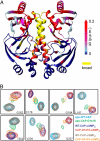
cAMP versus cGMP binding to CAP. Effect of (A) cAMP and (B) cGMP binding on the structure of CAP as assessed by chemical shift mapping. For direct comparison, chemical shift difference (Δδ) values, measured as described in ref. , are mapped by continuous-scale color onto the structure of CAP-cAMP2. (C) Superposition of the CAP-cAMP2 crystal structure (6) with the structural model of CAP-cGMP2 (this work). A close-up of the nucleotide-binding site is shown. Dotted lines denote hydrogen bonds between the nucleotide and CAP.
Fig. 5.
Fig. 5.
Effect of the CAP* G141S substitution on CAP. (A) The Δδ values are mapped by continuous-scale color onto the structure of CAP-cAMP2. Residues whose resonances broaden significantly are colored yellow. (B) Overlaid 1H-15N HSQC spectra of characteristic DBD residues of WT-CAP and CAP*-G141S in the apo, cAMP-bound, and cGMP-bound states.
Fig. 6.
Fig. 6.
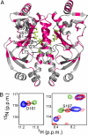
Effect of the CAP* T127L/S128I substitution on CAP. (A) Residues that experience significant chemical shift change as a result of the T127L/S128I double substitution are mapped (pink) on the structure of CAP-cAMP2. Effector domain residues that are involved in hydrophobic interactions with Ile-128, as assessed on the basis of chemical shift changes, are shown as green sticks. (B) Overlaid 1H-15N HSQC spectra of characteristic DBD residues of apo-CAP (blue), CAP-cAMP2 (green), and apo-CAP-T127L/S128I (red).
Fig. 7.
Fig. 7.
Mechanism of allosteric control of CAP. Schematic models of CAP in the 3 structurally characterized states: apo-CAP (this work), CAP-cAMP2 (6), and CAP-cAMP2-DNA (7). The proposed primary mechanism of allosteric control is clear and simple: cAMP binds to the CBD of CAP and makes direct contacts with Thr-127 and Ser-128. These contacts induce a coil-to-helix transition that extends the C-helix, and the intersubunit C-helix/C′-helix coiled coil, by 3 turns of helix. This coil-to-helix transition results in rotation of the DBDs of the CAP dimer by ≈60° and translation of the DBDs of the CAP dimer by ≈7 Å (distance of intersubunit F-helices is 41 Å in apo-CAP and 34 Å, matching the distance between successive DNA major grooves in CAP-cAMP2). This rotation and translation places the F-helices (“recognition helices”) of the DBDs of the CAP dimer in the correct orientation and correct position to interact with successive DNA major grooves. (See also Movie S1).

Comment in

References

    1. Lawson CL, et al. Catabolite activator protein: DNA binding and transcription activation. Curr Opin Struct Biol. 2004;14:10–20. - PMC - PubMed
    1. Martinez-Antonio A, Collado-Vides J. Identifying global regulators in transcriptional regulatory networks in bacteria. Current Opinion in Microbiology. 2003:482–489. - PubMed
    1. Benoff B, et al. Structural basis of transcription activation: The CAP-αCTD-DNA complex. Science. 2002;297:1562–1566. - PubMed
    1. Harman JG. Allosteric regulation of the cAMP receptor protein. Biochim Biophys Acta. 2001;1547:1–17. - PubMed
    1. McKay DB, Steitz TA. Structure of catabolite gene activator protein at 2.9 Å resolution suggests binding to left-handed B-DNA. Nature. 1981;290:744–749. - PubMed

Publication types

MeSH terms

Associated data