Comparative Study
. 2009 Jun;30(6):1016-23.
doi: 10.1093/carcin/bgp082. Epub 2009 Apr 9.

Lung tumor promotion by curcumin

Affiliations
Comparative Study

Lung tumor promotion by curcumin

Stephanie T Dance-Barnes et al. Carcinogenesis. 2009 Jun.

Erratum in

  • Carcinogenesis. 2010 Oct;31(10):1903. Dosage error in published abstract; MEDLINE/PubMed abstract corrected; Dosage error in article text

Abstract

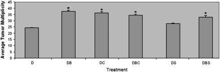
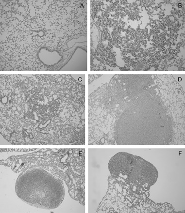
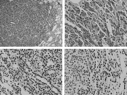
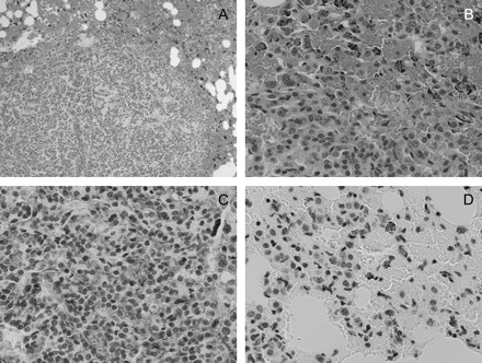
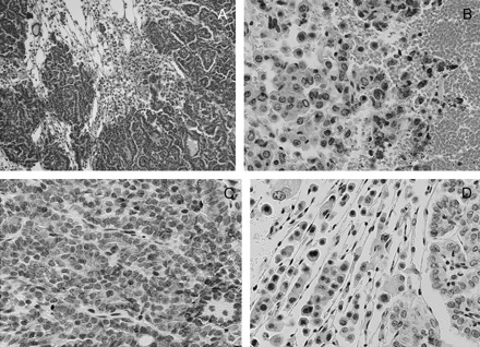
Curcumin exhibits anti-inflammatory and antitumor activity and is being tested in clinical trials as a chemopreventive agent for colon cancer. Curcumin's chemopreventive activity was tested in a transgenic mouse model of lung cancer that expresses the human Ki-ras(G12C) allele in a doxycycline (DOX) inducible and lung-specific manner. The effects of curcumin were compared with the lung tumor promoter, butylated hydroxytoluene (BHT), and the lung cancer chemopreventive agent, sulindac. Treatment of DOX-induced mice with dietary curcumin increased tumor multiplicity (36.3 +/- 0.9 versus 24.3 +/- 0.2) and progression to later stage lesions, results which were similar to animals that were co-treated with DOX/BHT. Microscopic examination showed that the percentage of lung lesions that were adenomas and adenocarcinomas increased to 66% in DOX/BHT, 66% in DOX/curcumin and 49% in DOX/BHT/curcumin-treated groups relative to DOX only treated mice (19%). Immunohistochemical analysis also showed increased evidence of inflammation in DOX/BHT, DOX/curcumin and DOX/BHT/curcumin mice relative to DOX only treated mice. In contrast, co-treatment of DOX/BHT mice with 200 p.p.m. [DOSAGE ERROR CORRECTED] of sulindac inhibited the progression of lung lesions and reduced the inflammation. Lung tissue from DOX/curcumin-treated mice demonstrated a significant increase (33%; P = 0.01) in oxidative damage, as assessed by the levels of carbonyl protein formation, relative to DOX-treated control mice after 1 week on the curcumin diet. These results suggest that curcumin may exhibit organ-specific effects to enhance reactive oxygen species formation in the damaged lung epithelium of smokers and ex-smokers. Ongoing clinical trials thus may need to exclude smokers and ex-smokers in chemopreventive trials of curcumin.


Figures

Fig. 1.
Fig. 1.
Average tumor multiplicity of mice at 45 weeks after the initiation of DOX treatment. The average tumor multiplicity for each treatment group is expressed as number of tumors per mouse/total number of mice with tumors ±SE. DOX only treated group (D), DOX/BHT co-treated group (DB), DOX/BHT/sulindac co-treated group (DBS), DOX/sulindac co-treated group (DS), DOX/BHT/curcumin co-treated group (DBC) and DOX/curcumin co-treated group (DC). *Denotes statistical significance from DOX-treated group, where P-value is <0.05.
Fig. 2.
Fig. 2.
Percent microscopic lesions grouped by tumor stage and treatment group. Percent proliferative lesions are expressed as the total number of lesions for each histological type within each treatment group/total number of proliferative lesions for each treatment group multiplied by 100 ± SE. The percentage for each microscopic lesion type is shown for hyperplasias (H), AD and large AD with atypical features and/or carcinomas (C) for the DOX (D), DOX/sulindac (DS), DOX/BHT (DB), DOX/BHT/sulindac (DBS), DOX/curcumin (DC) and DOX/BHT/curcumin (DBC) treatment groups. *Denotes statistical significance between hyperplasias (H), ** AD and *** carcinomas (C) when compared with the DOX-treated group.
Fig. 3.
Fig. 3.
Photomicrographs of mouse lung tissue. Hematoxylin and eosin stains of tissue from control and treated mice. (A) Normal lung with typically thin alveolar walls. (B) Diffuse pneumocyte hyperplasia in a mouse treated with DOX. Alveolar walls are thickened by plump, closely spaced pneumocytes. (C) Central focus of pneumocyte hyperplasia in a mouse treated with DOX. Alveolar walls are thickened about twice normal by plump, closely spaced pneumocytes. More normal lung is present adjacent. (D) Two solid coalescing AD in a mouse treated with DOX/BHT. Cells form dense sheets which expand the adjacent parenchyma. (E) Subpleural adenoma in a mouse treated with DOX/curcumin. The central area of pallor reflects necrosis. (F) Subpleural adenoma with papillary morphology in a mouse treated with DOX/BHT/curcumin. Panels A and B are ×10 and panels C–F are ×4 magnification.
Fig. 4.
Fig. 4.
Hematoxylin and eosin and immunohistochemical staining of lung tissue from DOX-treated mice. Panels (A) and (B) are ×10 and ×40 magnifications, respectively, of hematoxylin and eosin-stained slides showing relatively normal-appearing tissue with evidence of some hyperplasia. Panels (C) and (D) are immunohistochemical staining of lung tissue slides with antibodies to COX-2 and NF-κβ (Ser276) showing little staining for either protein in the tissues; magnification for IHC ×40.
Fig. 5.
Fig. 5.
Hematoxylin and eosin and immunohistochemical staining of lung tissue from DOX/BHT-treated mice. Panels (A) and (B) are ×10 and ×40 magnifications, respectively, of hematoxylin and eosin-stained slides showing mild edema around the tumor; panels (C) and (D) are immunohistochemical staining of lung tissue slides with antibodies to COX-2 and NF-κβ (Ser276) showing COX-2 and NF-κβ positivity in blood vessels and stromal cells, respectively; magnification for IHC ×40.
Fig. 6.
Fig. 6.
Hematoxylin and eosin and immunohistochemical staining of lung tissue from DOX/curcumin-treated mice. Panels (A) and (B) are ×10 and ×40 magnifications, respectively, of hematoxylin and eosin-stained slides showing marked inflammation around the tumors and a high density of red blood cells suggestive of edema (panel A) with infiltration of macrophages (panel B, cells with black dots in panel D). (C) Immunohistochemical staining of lung tissue slides with an antibody to COX-2. The tumor has a high-density vasculature that contains endothelial cells staining positively for COX-2. (D) Immunohistochemical staining of lung tissue slides with an antibody to NF-κβ (Ser276) demonstrating NF-κβ positive macrophages infiltrating the tumor tissue; magnification for IHC ×40.
Fig. 7.
Fig. 7.
Hematoxylin and eosin and immunohistochemical staining of lung tissue from DOX/BHT/curcumin-treated mice. Panels (A) and (B) are ×10 and ×40 magnifications, respectively, of hematoxylin and eosin-stained slides showing severe inflammation and leukocyte infiltration of the tumor. Panels (C) and (D) are immunohistochemical staining of lung tissue slides with antibodies to COX-2 and NF-κβ (Ser276) showing COX-2 and NF-κβ positivity in blood vessels, stromal and immune cells in the tumor microenvironment; magnification for IHC ×40.
Fig. 8.
Fig. 8.
Curcumin-mediated increase in oxidative damage in lung tissue. CCSP-rtTA/Ki-rasG12C bitransgenic mice were treated with 500 μg/ml of DOX in the drinking water. Two days after the initiation of DOX treatment, mice were kept on the normal diet or were fed diet supplemented with 4000 p.p.m. of curcumin. Nine days after the initiation of DOX treatment, the mice were euthanized and the lungs removed and homogenized in digestion buffer. Five microliters (0.13–2.2 mg) of supernatant were denatured and derivatized with 2,4-dinitrophenylhydrazine (DNPH), the derivatized proteins separated on 12% sodium dodecyl sulfate–polyacrylamide gel electrophoresis gels, transferred onto a nitrocellulose membrane and incubated with a primary antibody to either the DNP moiety of the protein or β-actin followed by a secondary horseradish peroxidase-conjugated antibody. The upper bands in the blot are non-specific binding that occurs when 2,4-dinitrophenylhydrazine-treated lysates are probed with the antibody to β-actin.

References

    1. Jemal A, et al. Cancer statistics, 2007. CA Cancer J. Clin. 2007;57:43–66. - PubMed
    1. Sandler AB, et al. COX-2 inhibition and lung cancer. Semin. Oncol. 2004;31:45–52. - PubMed
    1. Psaty BM, et al. Risks and benefits of celecoxib to prevent recurrent adenomas. N. Engl. J. Med. 2006;355:950–952. - PubMed
    1. Aggarwal BB, et al. Anticancer potential of curcumin: preclinical and clinical studies. Anticancer Res. 2003;23:363–398. - PubMed
    1. Duvoix A, et al. Chemopreventive and therapeutic effects of curcumin. Cancer Lett. 2005;223:181–190. - PubMed

Publication types

MeSH terms