. 2009 May 8;104(9):1049-57.
doi: 10.1161/CIRCRESAHA.108.190751. Epub 2009 Apr 9.

Antiinflammatory effects of the ETS factor ERG in endothelial cells are mediated through transcriptional repression of the interleukin-8 gene

Affiliations

Antiinflammatory effects of the ETS factor ERG in endothelial cells are mediated through transcriptional repression of the interleukin-8 gene

Lei Yuan et al. Circ Res. .

Abstract

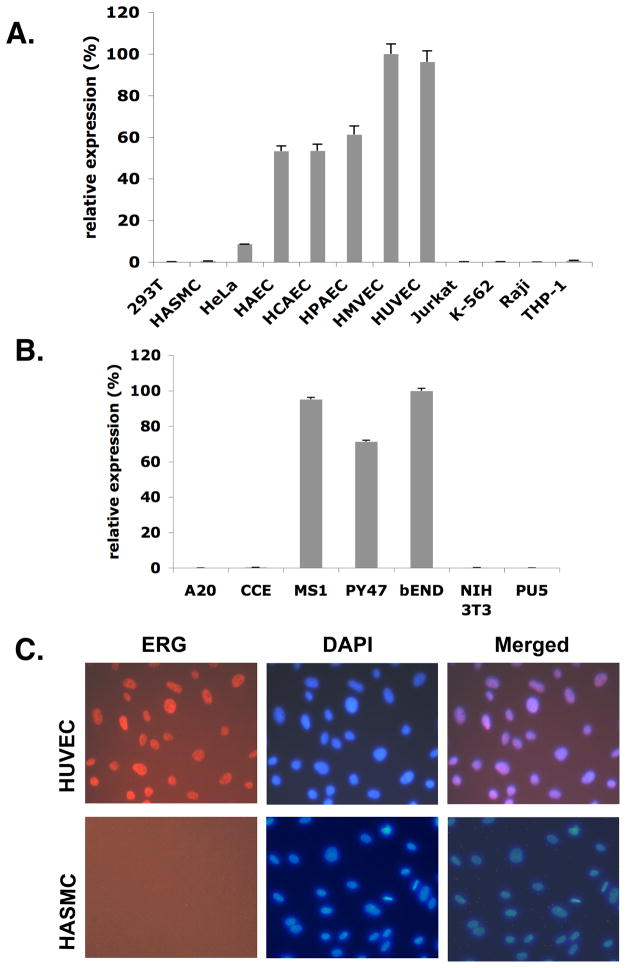
ERG (Ets-related gene) is an ETS transcription factor that has recently been shown to regulate a number of endothelial cell (EC)-restricted genes including VE-cadherin, von Willebrand factor, endoglin, and intercellular adhesion molecule-2. Our preliminary data demonstrate that unlike other ETS factors, ERG exhibits a highly EC-restricted pattern of expression in cultured primary cells and several adult mouse tissues including the heart, lung, and brain. In response to inflammatory stimuli, such as tumor necrosis factor-alpha, we observed a marked reduction of ERG expression in ECs. To further define the role of ERG in the regulation of normal EC function, we used RNA interference to knock down ERG. Microarray analysis of RNA derived from ERG small interfering RNA- or tumor necrosis factor-alpha-treated human umbilical vein (HUV)ECs revealed significant overlap (P<0.01) in the genes that are up- or downregulated. Of particular interest to us was a significant change in expression of interleukin (IL)-8 at both protein and RNA levels. Exposure of ECs to tumor necrosis factor-alpha is known to be associated with increased neutrophil attachment. We observed that knockdown of ERG in HUVECs is similarly associated with increased neutrophil attachment compared to control small interfering RNA-treated cells. This enhanced adhesion could be blocked with IL-8 neutralizing or IL-8 receptor blocking antibodies. ERG can inhibit the activity of the IL-8 promoter in a dose dependent manner. Direct binding of ERG to the IL-8 promoter in ECs was confirmed by chromatin immunoprecipitation. In summary, our findings support a role for ERG in promoting antiinflammatory effects in ECs through repression of inflammatory genes such as IL-8.


Figures

Figure 1
Figure 1. ERG expression in various human and mouse cells
RNAs were extracted from different cell types. Quantitative real-time PCR was performed using ERG-specific primers (n=3). GAPDH was used as an internal control. (A) ERG expression in human EC and non-EC. The results are shown as relative percentage compared with HMVEC. 293T--human fibroblast cell. HASMC--human aortic smooth muscle cell. HeLa--human epithelial cell. HAEC--human aortic EC. HCAEC--human coronary artery EC. HPAEC--human pulmonary artery EC. HMVEC--human microvascular EC. HUVEC--human umbilical vein EC. Jurkat--human T cell. K-562--human erythroid leukemia cell. Raji--human B cell. THP 1-- human monocyte. (B) ERG expression in mouse EC and non-EC. The results are shown as relative percentage compared with bEND. A20—murine B cell. MS1—murine EC. PY41—murine EC. bEND—cerebellar EC. NIH 3T3—murine fibroblast cell. PU5—murine T cell. (C) Immunofluorescent staining of ERG in cells. Cells were formalin fixed and stained with anti-ERG antibody (red). Nuclei were stained by DAPI (blue).
Figure 2
Figure 2. Examination of ERG expression in mouse tissues
(A) RNAs were extracted from a variety of mouse tissues. cDNA was prepared from RNAs and utilized in quantitative real-time PCR. ERG expression was analyzed using ERG-specific primers and normalized against GAPDH (n=3). The data are shown as relative percentage compared with heart. (B) Representative images of immunofluorescent staining of ERG in mouse brain and heart. Tissues were frozen-sectioned and stained for ERG (green), VE-cadherin (red), or DAPI (blue). Magnification X40. Scale bar=75 mm.
Figure 3
Figure 3. Regulation of inflammatory cytokines on ERG protein level
(A,B) HUVEC were initially cultured in 6-well plates in serum-free medium for 12 hours and then stimulated with 2 ng/ml TNF-α (A) or10 ng/ml LPS (B) for the periods of time indicated. Extracts were prepared and equal amounts of total proteins were separated on polyacrylamide gel. Protein levels of ERG and the loading marker tubulin were determined by Western blot analysis. Representative Western blots show protein bands of ERG and tubulin (A and B, top). Densitometry analysis shows density ratios of ERG and tubulin (A and B, bottom). Three experiments were performed. * P values <0.01. (C, D) Representative images of ERG immunofluorescent stain of heart tissues from mouse models of endotoxemia. Heart tissues were harvested at 24 hours after LPS administration or CLP. Formalin-fixed cryosections were used for double immunofluorescent stain for CD31 (red) and ERG (green). Scale bar=75mm.
Figure 4
Figure 4. Effect of ERG on neutrophil attachment
(A, B) Effect of ERG siRNA transfection on ERG expression. HUVEC were transfected with ERG siRNA (40 nmol/L) or a scrambled control siRNA (40 nmol/L) as a control. ERG expression was assessed by Western blot using anti-ERG antibody (A) or by quantitative real-time PCR using ERG-specific primers (B) normalized against GAPDH (n=3). * P < 0.01. (C, D) ERG siRNA- or control siRNA-treated HUVEC were cultured in medium alone or 2 ng/ml TNF-α as indicated for additional 12 hours. Neutrophils were isolated freshly from human blood samples and incubated with HUVEC. Cells were washed 3X with PBS before imaging. The number of adherent neutrophils was assessed in five randomly selected fields for each condition in triplicate samples. The data are shown as fold change in comparison with control siRNA treated samples (C). * P < 0.01. Representative phase-contrast images of corresponding treatments are shown (D).
Figure 5
Figure 5. Gene Expression based comparison of ERG siRNA and TNF-α stimulated HUVEC
(A) Venn diagram indicating overlap of differentially expressed genes between ERG siRNA and TNF-α stimulated HUVEC. (B) Heatmap indicating expression of overlapping genes (shown in Venn diagram) in the ERG siRNA transfected HUVEC. (C) Heatmap indicating expression of overlapping genes in the TNF-α stimulated HUVEC. The columns represent the samples and rows represent the genes. Gene expression is shown with pseudocolor scale (−3 to 3) with red denoting high expression level and green denoting low expression level of gene.
Figure 6
Figure 6. Identification of novel downstream targets of ERG
HUVEC were transfected with either ERG siRNA (40 nmol/L) or a control siRNA (40 nmol/L). After 48 hours of incubation: (A) cDNA was prepared from isolated RNA and analyzed by Q-PCR using primers corresponding to each gene indicated, normalized against GAPDH. The data are shown as fold change compared with control siRNA treated samples (n=3). (B, C) The supernatants were collected and assayed for the protein expression levels of genes indicated. It is represented as a fold change in comparison with control siRNA samples (B), or as the protein level in ng/ml (C) (n=3). (D) Effect of ERG supression on leukocyte adhesion is IL-8 dependent. ERG siRNA- or control siRNA-treated HUVEC were used for neutrophil attachment assays (as described in Fig. 4) in the presence of blocking antibodies to IL-8, IL-8 receptor (CXCR1 and/or CXCR2), or isotype-matched control normal IgG. Data are represented as fold-changes compared with control siRNA-treated cells. * P < 0.05.
Figure 7
Figure 7. IL-8 is a downstream target of ERG
(A) Schematic diagram of ERG binding sites. The 1.4 kb upstream promoter region of IL-8 was analyzed in search for the putative ERG binding site based on the alignment of ERG binding site derived from previously characterized ERG target genes. Red boxes indicate the putative ERG binding sites. The bidirectional arrows marked the target regions for ChIP assays (ChIP1 and ChIP2). (B) ChIP assay of IL-8 promoter using HUVEC. An ERG polyclonal antibody was used for precipitation. PCR analysis of the input, in the absence of ERG antibody (CTR), and in the presence of ERG antibody (ERG) after immunoprecipitation (IP) using primers corresponding to two ERG putative binding sites (ChIP1 and ChIP2) of the IL-8 promoter. Molecular weight markers are shown on the left. (C, D) Transactivation assay. HUVEC were co-transfected with IL-8 promoter (IL-8 wt) or IL-8 promoter containing mutation at ChIP2 (IL-8 mut) in reporter gene vector and pCI expressing vector encoding ERG or empty vector. After 24 hours of incubation, cells were collected for luciferase assay. The data are shown as relative luciferase activity (%) compared with cotransfection with empty expression plasmid (n=3). ** P<0.05. * P < 0.01.

References

    1. Wasylyk B, Hahn SL, Giovane A. The Ets family of transcription factors. Eur J Biochem. 1993;211:7–18. - PubMed
    1. Gu X, Shin BH, Akbarali Y, Weiss A, Boltax J, Oettgen P, Libermann TA. Tel-2 is a novel transcriptional repressor related to the Ets factor Tel/ETV-6. J Biol Chem. 2001;276:9421–9436. - PubMed
    1. Kas K, Finger E, Grall F, Gu X, Akbarali Y, Boltax J, Weiss A, Oettgen P, Kapeller R, Libermann TA. ESE-3, a novel member of an epithelium-specific ets transcription factor subfamily, demonstrates different target gene specificity from ESE-1. J Biol Chem. 2000;275:2986–2998. - PubMed
    1. Oettgen P, Finger E, Sun Z, Akbarali Y, Thamrongsak U, Boltax J, Grall F, Dube A, Weiss A, Brown L, Quinn G, Kas K, Endress G, Kunsch C, Libermann TA. PDEF, a novel prostate epithelium-specific ets transcription factor, interacts with the androgen receptor and activates prostate-specific antigen gene expression. J Biol Chem. 2000;275:1216–1225. - PubMed
    1. Oettgen P, Akbarali Y, Boltax J, Best J, Kunsch C, Libermann TA. Characterization of NERF, a novel transcription factor related to the Ets factor ELF-1. Mol Cell Biol. 1996;16:5091–5106. - PMC - PubMed

Publication types

MeSH terms