

Regulation of signaling pathways downstream of IGF-I/insulin by androgen in skeletal muscle of glucocorticoid-treated rats
- PMID: 19359918
- PMCID: PMC4085982
- DOI: 10.1097/TA.0b013e31817e7420
Regulation of signaling pathways downstream of IGF-I/insulin by androgen in skeletal muscle of glucocorticoid-treated rats
Abstract
Background: The mechanisms by which androgens ameliorate glucocorticoid-induced muscle wasting are still under investigation. In the present study, we tested the hypothesis that androgen's effects in reversing muscle wasting are related to activating the signaling pathways downstream of insulin-like growth factor-1 (IGF-I)/insulin.
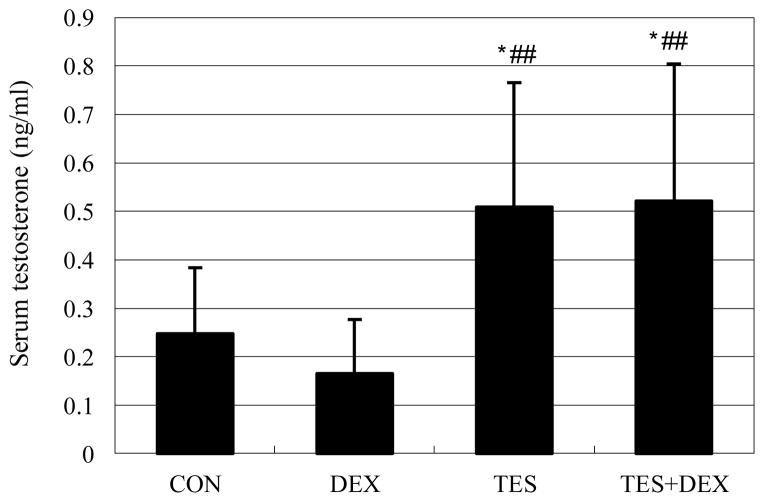
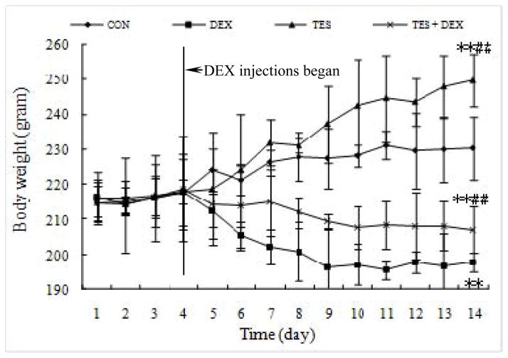
Methods: Forty female Sprague-Dawley rats were randomly divided into four groups: control group, dexamethasone (DEX) group, testosterone (TES) group, and TES + DEX group. Each group was injected with saline or DEX (0.1 mg/100 g/d) for 10 days and sesame oil or TES (0.5 mg/100 g/d) for 13 days. Several downstream targets of IGF-I/insulin in skeletal muscle including protein kinase B (Akt), p70 ribosomal protein S6 kinase (p70S6K), and glycogen synthase kinase-3beta (GSK-3beta) that are associated with protein synthesis were examined. Two proteolysis-related ubiquitin E3-ligases, muscle atrophy F-box, and muscle RING finger-1 that are also regulated by IGF-I/insulin were also assessed.
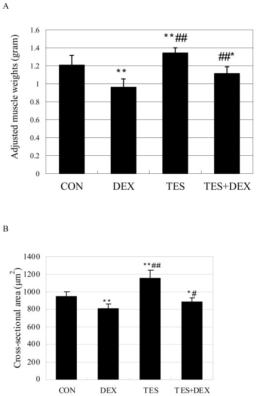
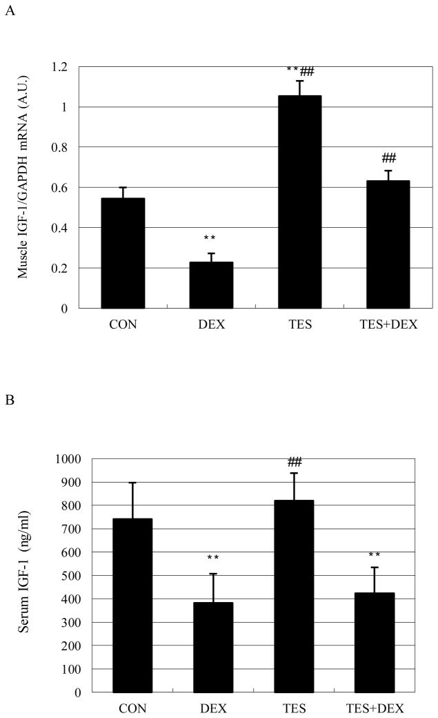
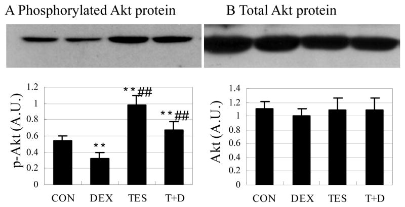
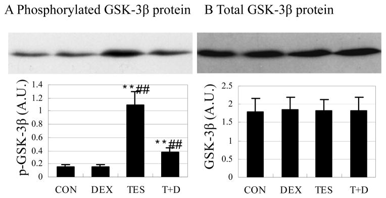
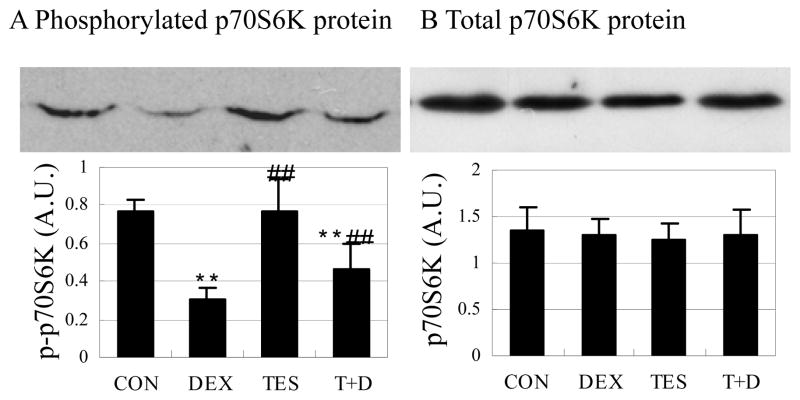
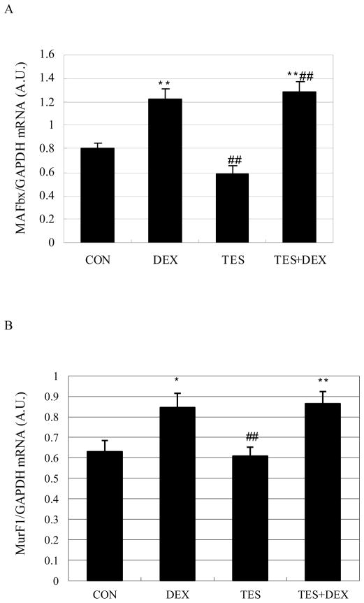
Results: TES attenuated gastrocnemius muscle atrophy induced by DEX. TES prevented the DEX-induced decrease of IGF-I expression in gastrocnemius muscle, but not in serum. TES ameliorated DEX-induced dephosphorylation of Akt and p70S6K and promoted the phosphorylation of GSK-3beta in gastrocnemius muscle. The total amount of Akt, p70S6K, or GSK-3beta proteins was not changed among these groups. TES did not show any effects on the DEX-induced upregulation of muscle atrophy F-box, and muscle RING finger-1 mRNA in gastrocnemius muscle.
Conclusion: This findings suggest that the effects of TES in reversing DEX-induced muscle atrophy are related to signaling pathways downstream of IGF-I/insulin that are associated with protein synthesis.
Figures
References
-
- Fang CH, James HJ, Ogle C, Fischer JE, Hasselgren PO. Influence of burn injury on protein metabolism in different types of skeletal muscle and the role of glucocorticoids. J Am Coll Surg. 1995;180:33–42. - PubMed
-
- Crawford BA, Liu PY, Kean MT, Bleasel JF, Handelsman DJ. Randomized placebo-controlled trial of androgen effects on muscle and bone in men requiring long-term systemic glucocorticoid treatment. J Clin Endocrinol Metab. 2003;88:3167–3176. - PubMed
-
- Creutzberg EC, Schols AM. Anabolic steroids. Curr Opin Clin Nutr Metab Care. 1999;2:243–253. - PubMed
-
- Creutzberg EC, Wouters EF, Mostert R, Pluymers RJ, Schols AM. A role for anabolic steroids in the rehabilitation of patients with COPD? A double-blind, placebo-controlled, randomized trial. Chest. 2003;124:1733–1742. - PubMed
Publication types
MeSH terms
Substances
Grants and funding
LinkOut - more resources
Full Text Sources
Medical
Molecular Biology Databases
