. 2009;4(4):e5181.
doi: 10.1371/journal.pone.0005181. Epub 2009 Apr 10.

Newly generated heparanase knock-out mice unravel co-regulation of heparanase and matrix metalloproteinases

Affiliations

Newly generated heparanase knock-out mice unravel co-regulation of heparanase and matrix metalloproteinases

Eyal Zcharia et al. PLoS One. 2009.

Abstract

Background: Heparanase, a mammalian endo-beta-D-glucuronidase, specifically degrades heparan sulfate proteoglycans ubiquitously associated with the cell surface and extracellular matrix. This single gene encoded enzyme is over-expressed in most human cancers, promoting tumor metastasis and angiogenesis.

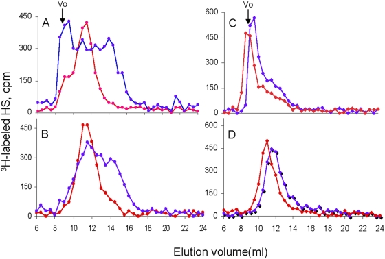
Principal findings: We report that targeted disruption of the murine heparanase gene eliminated heparanase enzymatic activity, resulting in accumulation of long heparan sulfate chains. Unexpectedly, the heparanase knockout (Hpse-KO) mice were fertile, exhibited a normal life span and did not show prominent pathological alterations. The lack of major abnormalities is attributed to a marked elevation in the expression of matrix metalloproteinases, for example, MMP2 and MMP14 in the Hpse-KO liver and kidney. Co-regulation of heparanase and MMPs was also noted by a marked decrease in MMP (primarily MMP-2,-9 and 14) expression following transfection and over-expression of the heparanase gene in cultured human mammary carcinoma (MDA-MB-231) cells. Immunostaining (kidney tissue) and chromatin immunoprecipitation (ChIP) analysis (Hpse-KO mouse embryonic fibroblasts) suggest that the newly discovered co-regulation of heparanase and MMPs is mediated by stabilization and transcriptional activity of beta-catenin.

Conclusions/significance: The lack of heparanase expression and activity was accompanied by alterations in the expression level of MMP family members, primarily MMP-2 and MMP-14. It is conceivable that MMP-2 and MMP-14, which exert some of the effects elicited by heparanase (i.e., over branching of mammary glands, enhanced angiogenic response) can compensate for its absence, in spite of their different enzymatic substrate. Generation of viable Hpse-KO mice lacking significant abnormalities may provide a promising indication for the use of heparanase as a target for drug development.


Conflict of interest statement

Competing Interests: The authors have declared that no competing interests exist.

Figures

Figure 1
Figure 1. Targeted disruption of the heparanase gene and generation of heparanase deficient mice.
A. Structure of the 5′ end of the heparanase (Hpse) gene (top-normal allele), the targeting vector (middle- K/O allele), and the expected size of products obtained by digestion with Sca1 or EcoRv (bottom). The orientation of the neo cassette and the southern blot probes are indicated. B. Southern blot analysis. Genomic DNA was extracted from embryos of the intercross of Hpse +/− heterozygous mice and subjected to Southern blot analysis after digestion with ERv. Wild type (wt) embryos exhibited only the normal allele, heterozygous embryos exhibited both the normal and the mutated allele, while Hpse-KO mice exhibited only the shorter, KO allele. Samples were hybridized with the external probe 3, shown in A. C. Heparanase mRNA expression. RNA was extracted from lungs and spleens of wt and Hpse-KO mice and subjected to PCR analysis of heparanase expression using 3 different PCR primer pairs designed to amplify different regions of the Hpse gene, as indicated. Heparanase expression was identified in samples derived from wt but not from Hpse-KO mice. D. Heparanase activity. Blood samples derived from 4 wt and 4 Hpse-KO mice, as well as total cell lysate derived from JAR cells, were incubated (16 h, 37°C, pH 6.2) with sulfate labeled ECM. Labeled degradation products released into the incubation medium were subjected to gel filtration analysis on Sepharose 6B, as described in “Materials and Methods”. High heparanase activity was noted only in samples derived from wt mice; no heparanase activity was detected in Hpse-KO samples or JAR cell lysate. E. HS degradation. Liver, kidney and spleen tissue extracts derived from wt and Hpse-KO mice were prepared as described in “Materials and Methods” and incubated (18 h, 37°C, pH 5.8) with 3H-acetyl labeled HS. The reaction mixture was then subjected to gel chromatography on Superose-12. The upper panel shows blank (peak I; control 3H-acetyl labeled HS substrate) and positive control (control substrate treated with recombinant heparanase; peak II). Incubations of Hpse-KO tissue extracts (black) resulted in the same elution profile as the blank incubation (upper panel), indicating no detectable heparanase activity. In contrast, incubations with wt tissue extracts (purple) resulted in substantial cleavage of the HS substrate, similar to incubation with recombinant heparanase (upper panel).
Figure 2
Figure 2. Molecular structure of HS from wt vs. Hpse-KO mice.
Total metabolically 35S-labeled HSPGs was isolated as described in “Materials and Methods”. The samples were analyzed on a Superose 12 column as shown for liver (A) and kidney (C). The HSPGs were treated with alkali and the released free HS chains were analyzed on the same column as show for liver (B) and kidney (D). (Blue- wt; red- Hpse-KO). Standard heparin is eluted at a volume of 14 ml.
Figure 3
Figure 3. Morphological appearance of mammary glands from wt vs. Hpse-KO mice.
Whole-mount preparations of mammary glands from 3-month-old virgin mice were stained with hematoxylin. Hpse-KO derived mammary glands (right panel) showed abundant side branches and alveolar structures compared with glands from age-matched wt animals (left panel).
Figure 4
Figure 4. Endothelial sprouting and angiogenesis.
A. FGF-2 induced vascular sprouting in the aortic ring model. Hpse-KO and wt derived aortic rings were subjected to FGF-2 induced vascular sprouting for 6 days. The rings were then fixed, stained with 0.02% crystal violet and evaluated for vascular sprouting. A more extensive endothelial sprouting was noted in Hpse-KO derived rings (right panel) as compared to wt derived rings (left panel). B. Matrigel plug assay. Hpse-KO (lower panels) and wt (upper panels) mice were injected subcutaneously with 200 µl of growth factor depleted Matrigel supplemented with FGF-2 (80 ng/ml). Seven days later, the Matrigel plugs were excised and photographed, followed by homogenization and determination of hemoglobin content using Drabkin's reagent (right). A pronounced angiogenic response was noted in Hpse-KO vs. wt mice (55±7.18 mg/dl vs. 21±6.2 mg/dl; p≤0.0002, respectively).
Figure 5
Figure 5. Hpa2 expression.
RNA samples derived from liver, kidney and mammary glands of wt and Hpse-KO mice were subjected to quantitative real time PCR analysis to evaluate the expression of Hpa2. The expression level determined for each MMP in the wt tissue (blue) was regarded as 100% and the corresponding expression level determined in the Hpse-KO tissue (purple) is presented as percentage relative to the 100% value. Each reaction was repeated 6 times and the mean±SD is indicated. No significant difference was detected in Hpa2 expression between wt and Hpse-KO mice.
Figure 6
Figure 6. MMP expression in Hpse-KO mice.
A. Real-time PCR. RNA was extracted from liver, kidney and mammary gland of wt and Hpse-KO mice and subjected to quantitative real time PCR analysis to evaluate the expression of MMP-2, MMP-9, MMP-14 and MMP-25. The expression level determined for each MMP in the wt tissue (blue) was regarded as 100% and the corresponding expression determined in the Hpse-KO tissue (Purple) are presented as percentage relative to it. Each reaction was repeated 6 times and the mean±SD is indicated. B. Western blot analysis. Liver, kidney and mammary gland tissue extracts, prepared as described in “Materials and Methods”, were subjected to Western blot analysis using anti-mouse MMP-2 monoclonal antibodies (mA801B; upper panels), anti mouse β-catenin (mAb610154; middle panels), or anti mouse α-tubulin (B-5-1-2; lower panels). Higher protein levels of MMP-2 and β-catenin were detected in samples derived from Hpse-KO vs. wt tissues. C. MMP2 zymography. Serum samples derived from Hpse-KO and wt mice were evaluated for MMP-2 activity. MMP-2 activity was approximately 3 fold higher in plasma samples derived from Hpse-KO vs. wt mice. D. β-catenin immunostaining. Parafin embedded kidney tissue sections were subjected to immunostaining with antibody directed against β-catenin. Increased staining was observed in kidney derived from Hpse-KO vs. wt mice. C. MMP2 zymography. Serum samples derived from Hpse-KO and wt mice were evaluated for MMP-2 activity. MMP-2 activity was approximately 3 fold higher in plasma samples derived from Hpse-KO vs. wt mice.
Figure 7
Figure 7. MMP expression and ChIP analysis for β-catenin in MEF cells derived from Hpse-KO and wt mice.
A. Real-time PCR. RNA was extracted from MEF derived from wt and Hpse-KO mice and subjected to quantitative real time PCR analysis to evaluate the expression of MMP-2, MMP-9, and MMP-14. The expression level determined for each MMP in the wt tissue (blue) was regarded as 100% and the corresponding levels determined in the Hpse-KO cells (purple) are presented as percentage relative to it. Each reaction was repeated 6 times and the mean±SD is indicated. B. Schematic representation of regions along the murine MMP-14 promoter with ≥95% homology to the consensus Lef/Tcf motif. Numbers show the location of putative Lef/Tcf motifs relative to the transcription initiation site (bent arrow). C. ChIP analysis. Following cross-linking of proteins to DNA, chromatin derived from Hpse-KO and wt MEF was sonicated into fragments of average length ≤500 bp; the β-catenin protein was immunoprecipitated with anti β-catenin antibody, and PCR analysis was performed on the immunoprecipitated DNA samples using primer sets MMP14-Pro1 & Pro2, as described in ‘Materials and Methods’. Samples were equilibrated for DNA loading amounts using primers specific to actin. The results are representative of three independent experiments.
Figure 8
Figure 8. Expression of MMPs in heparanase transfected MDA-231 human breast carcinoma cells.
MDA-MB-231 cells were transfected with a mock (empty vector) or either active (Hpse) or mutated inactive (Mut) heparanase gene. Heparanase (A) and MMPs (B) mRNA expression levels were determined by real-time PCR, as described under “Materials and Methods”. The appropriate primers are listed in Table 2. The expression levels determined in the mock transfected cells were regarded as 100%, and the levels in Hpse and mut-Hpse transfected cells were presented as percentage relative to the mock transfected cells. Decreased levels of MMP-2, MMP-9, and MMP-14 mRNAs were noted in cells over-expressing the active form of heparanase, but not the double mutant, inactive form of the enzyme.

References

    1. Bernfield M, Gotte M, Park PW, Reizes O, Fitzgerald ML, et al. Functions of cell surface heparan sulfate proteoglycans. Annu Rev Biochem. 1999;68:729–777. - PubMed
    1. Iozzo RV, San Antonio JD. Heparan sulfate proteoglycans: heavy hitters in the angiogenesis arena. J Clin Invest. 2001;108:349–355. - PMC - PubMed
    1. Kjellen L, Lindahl U. Proteoglycans: structures and interactions [published erratum appears in Annu Rev Biochem 1992;61:following viii]. Annu Rev Biochem. 1991;60:443–475. - PubMed
    1. Vlodavsky I, Bar-Shavit R, Korner G, Fuks Z. Extracellular matix-bound growth factors, enzymes and plasma proteins. In: Rohrbach DH, Timpl R, editors. Academic press Inc., editor. Basement membranes: Cellular and molecular aspects. Orlando, , Fl: 1993. pp. 327–343.
    1. Vlodavsky I, Folkman J, Sullivan R, Fridman R, Ishai-Michaeli R, et al. Endothelial cell-derived basic fibroblast growth factor: synthesis and deposition into subendothelial extracellular matrix. Proc Natl Acad Sci U S A. 1987;84:2292–2296. - PMC - PubMed

Publication types

MeSH terms