. 2009 Apr;5(4):e1000379.
doi: 10.1371/journal.ppat.1000379. Epub 2009 Apr 10.

The NOD/RIP2 pathway is essential for host defenses against Chlamydophila pneumoniae lung infection

Affiliations

The NOD/RIP2 pathway is essential for host defenses against Chlamydophila pneumoniae lung infection

Kenichi Shimada et al. PLoS Pathog. 2009 Apr.

Erratum in

  • PLoS Pathog. 2009 Apr;5(4). doi: 10.1371/annotation/0f4b8a96-1598-4c90-b816-8fbe7614310b
  • PLoS Pathog. 2009 Apr;5(4). doi: 10.1371/annotation/f3aa682e-3bc2-4a05-ac7f-05c6cfe1bbd7

Abstract

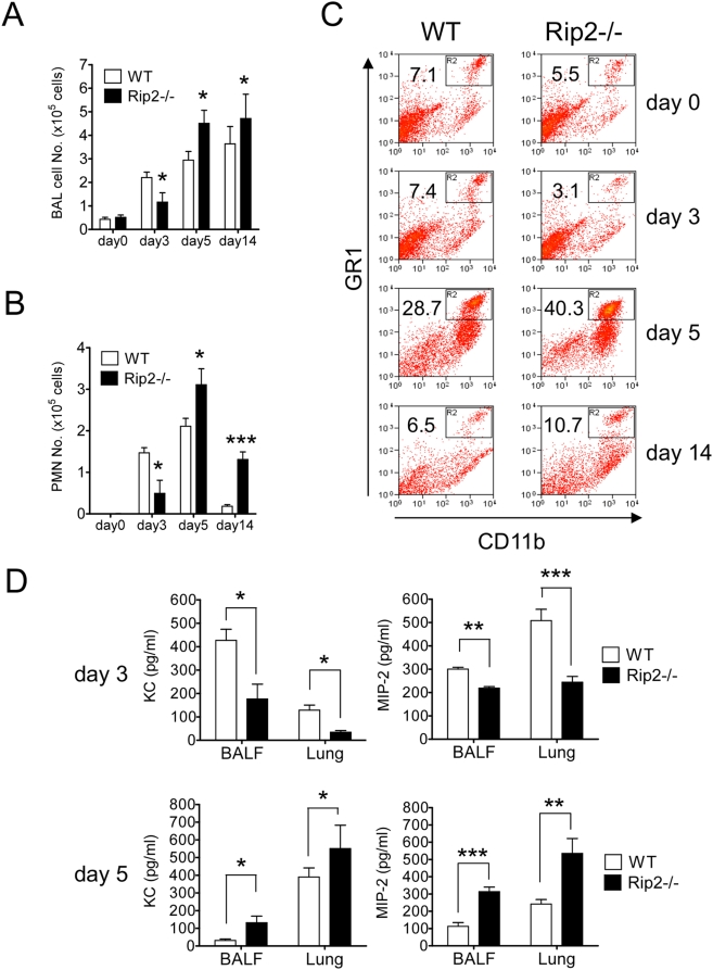
Here we investigated the role of the Nod/Rip2 pathway in host responses to Chlamydophila pneumoniae-induced pneumonia in mice. Rip2(-/-) mice infected with C. pneumoniae exhibited impaired iNOS expression and NO production, and delayed neutrophil recruitment to the lungs. Levels of IL-6 and IFN-gamma levels as well as KC and MIP-2 levels in bronchoalveolar lavage fluid (BALF) were significantly decreased in Rip2(-/-) mice compared to wild-type (WT) mice at day 3. Rip2(-/-) mice showed significant delay in bacterial clearance from the lungs and developed more severe and chronic lung inflammation that continued even on day 35 and led to increased mortality, whereas WT mice cleared the bacterial load, recovered from acute pneumonia, and survived. Both Nod1(-/-) and Nod2(-/-) mice also showed delayed bacterial clearance, suggesting that C. pneumoniae is recognized by both of these intracellular receptors. Bone marrow chimera experiments demonstrated that Rip2 in BM-derived cells rather than non-hematopoietic stromal cells played a key role in host responses in the lungs and clearance of C. pneumoniae. Furthermore, adoptive transfer of WT macrophages intratracheally was able to rescue the bacterial clearance defect in Rip2(-/-) mice. These results demonstrate that in addition to the TLR/MyD88 pathway, the Nod/Rip2 signaling pathway also plays a significant role in intracellular recognition, innate immune host responses, and ultimately has a decisive impact on clearance of C. pneumoniae from the lungs and survival of the infectious challenge.


Conflict of interest statement

The authors have declared that no competing interests exist.

Figures

Figure 1
Figure 1. Rip2−/− mice develop a severe and chronic lung inflammation and manifest impaired early cytokine production in response to C. pneumoniae infection.
WT and Rip2−/− mice were infected with C. pneumoniae (1×10⁶ IFU/mouse). Lungs of infected mice were isolated on days 3, 5, 14, and 35 after infection. Lungs were fixed in 10% buffered formalin, sectioned, and stained with H&E to determine the extent of inflammation (A). Histological analysis of inflammation in C. pneumoniae–infected lungs of WT and Rip2−/− mice on days 3, 5, 14, and 35 post-infection (B). Statistical significance was determined by Student's t test (*p<0.05, **p<0.01, ***p<0.001). At 3, 5, and 14 days post-infection, the levels of IL-6, IL-12p40, and IFN-γ in BALF and lung tissue homogenates were determined (C). Statistical significance was determined by Student's t test (*p<0.05, n = 8–14).
Figure 2
Figure 2. Delayed clearance of C. pneumoniae and increased mortality in Rip2−/− mice.
WT and Rip2−/− mice were infected with C. pneumoniae (1×10⁶ IFU/mouse). At 3, 5, and 14 days post-infection, the bacterial burden in lung tissue homogenates was determined (A). Statistical significance was determined by Student's t test (*p<0.05, **p<0.01). Mortality was monitored after infection (B). Statistical significance was determined by Fisher's exact test (*p<0.05, n = 11).
Figure 3
Figure 3. Rip2 deficiency impairs early neutrophil recruitment and chemokine expression in the lungs during C. pneumoniae infection.
C. pneumoniae was inoculated intratracheally into Rip2−/− and WT mice (1×10⁶ IFU/mouse). BALF was collected from WT and Rip2−/− mice on days 0, 3, 5, and 14 post-infection. Total BAL cells (A) and PMN (B) were counted. Statistical significance was determined by Student's t test (*p<0.05, ***p<0.001, n = 10∼14). Lungs were removed, and digested with collagenase and DNase I. Cells were stained with PE-anti-GR1 mAB and PECy5-anti-CD11b mAb and analyzed by FACScan (C). The percentages of gated positive cells are indicated. Data shown are representative of four independent experiments. The levels of KC and MIP-2 in BALF and lung tissue homogenates on days 3 and 5 post-infection were determined (D). Statistical significance was determined by Student's t test (*p<0.05, **p<0.01, ***p<0.001).
Figure 4
Figure 4. Identification of cell types in the lung harboring C. pneumoniae.
Single cell suspensions of infected lungs (day 5) were stained with FITC-anti-Chlamydia mAb and analyzed by flow cytometry to determine which cell types contained C. pneumoniae (A). Composition of lung leukocytes in infected mice (day 5) (B). Statistical significance was determined by Student's t test (*p<0.05, **p<0.01). Representative histograms were shown to analyze relative bacterial number in macrophages (MAC) and neutrophils (Neu) between WT (thin line histogram) and Rip2−/− (gray-filled histogram) (C).
Figure 5
Figure 5. Impaired iNOS mRNA expression and NO production in Rip2−/− mice.
Lung tissues of WT and Rip2−/− mice were removed at days 0, 1, and 5 post-infection. iNOS mRNA expression was measured by semi-quantitative RT-PCR (A). Bone marrow–derived macrophages (BMDM) were stimulated with C. pneumoniae for 24h. NO production was evaluated by measuring nitrite accumulation in the supernatant using the Griess reaction (B). Statistical significance was determined by Student's t test (*p<0.01, **p<0.001, n = 4). C. pneumoniae (MOI 5)–infected BMDMs were cultured in the presence of 5mM L-NMMA or D-NMMA for 5 days. Cell lysates were harvested and viable bacteria were quantified by infecting HEp2 cells (C). Statistical significance was determined by one-way ANOVA with Tukey's post-hoc test (*p<0.05, n = 4). Data shown are representative of two independent experiments.
Figure 6
Figure 6. Chemokine and NO production in Nod1−/− and Nod2−/− BMDM and quantitative bacterial counts in lungs of Nod1−/− and Nod2−/− mice following infection with C. pneumoniae.
WT, Nod1−/−, and Nod2−/− BMDMs were infected with C. pneumoniae for 24 hours, and KC (A) and NO (B) concentrations in the supernatant were determined. Data shown are representative of three independent experiments. Statistical significance was determined by Student's t test (*p<0.05, **p<0.01, n = 3–4). C. pneumoniae (MOI 5)–infected BMDM were cultured for 5 days. Cell lysates were harvested and viable bacteria were quantified by infecting HEp2 cells (C). Data shown are representative of two independent experiments. Statistical significance was determined by Student's t test (*p<0.05, n = 4). Nod1−/− and Nod2−/− mice were infected with C. pneumoniae (1×10⁶ IFU). After 5 days, lungs were collected and the quantitative bacterial counts in lung tissue homogenates were determined (D). Statistical significance was determined by Student's t test (*p<0.05, **p<0.01, n = 7).
Figure 7
Figure 7. Rip2 in BM-derived cells contributes to bacterial clearance from the lungs.
BM chimeric mice were created by lethally irradiating the recipient mice, then performing adoptive transfer and reconstitution with donor BM obtained form WT or Rip2−/− donor mice. After 7 weeks, BM-reconstituted mice were infected with C. pneumoniae (1×10⁶ IFU). Lungs were collected on day 5 post-infection. The bacterial burden in lung tissue homogenates was determined (A). BMDMs were grown and isolated from WT or Rip2−/− mice and adoptively transferred intratracheally into the airways of WT mice or Rip2−/− mice (5×10⁵/mouse) together with C. pneumoniae (1×10⁶ IFU). BMDMs were first incubated with C. pneumoniae in vitro for 30 minutes prior to intratracheal administration. After 5 days, lungs were collected and the bacterial burden in lung tissue homogenates was determined (B). Statistical significance was determined by one-way ANOVA with Tukey's post-hoc test (*p<0.05, n = 3–5).

Similar articles

Cited by

References

    1. Karvonen M, Tuomilehto J, Pitkäniemi J, Naukkarinen A, Saikku P. Chlamydia pneumoniae IgG antibody prevalence in south-western and eastern Finland in 1982 and 1987. Int J Epidemiol. 1994;23:176–184. - PubMed
    1. Campbell LA, Kuo CC. Chlamydia pneumoniae—an infectious risk factor for atherosclerosis? Nat Rev Mirobiol. 2004;2:23–32. - PubMed
    1. Gupta S, Leatham EW. The relation between Chlamydia pneumoniae and atherosclerosis. Heart. 1997;77:7–8. - PMC - PubMed
    1. Hansbro PM, Beagley KW, Horvat JC, Gibson PG. Role of atypical bacterial infection of the lung in predisposition/protection of asthma. Pharmacol Ther. 2004;101:193–210. - PubMed
    1. Belland RJ, Ouellette SP, Gieffers J, Byrne GI. Chlamydia pneumoniae and atherosclerosis. Cell Microbiol. 2004;6:117–127. - PubMed

Publication types

MeSH terms

Substances