. 2009 Apr;5(4):e1000374.
doi: 10.1371/journal.ppat.1000374. Epub 2009 Apr 10.

The Mycobacterium tuberculosis phagosome is a HLA-I processing competent organelle

Affiliations

The Mycobacterium tuberculosis phagosome is a HLA-I processing competent organelle

Jeff E Grotzke et al. PLoS Pathog. 2009 Apr.

Abstract

Mycobacterium tuberculosis (Mtb) resides in a long-lived phagosomal compartment that resists maturation. The manner by which Mtb antigens are processed and presented on MHC Class I molecules is poorly understood. Using human dendritic cells and IFN-gamma release by CD8(+) T cell clones, we examined the processing and presentation pathway for two Mtb-derived antigens, each presented by a distinct HLA-I allele (HLA-Ia versus HLA-Ib). Presentation of both antigens is blocked by the retrotranslocation inhibitor exotoxin A. Inhibitor studies demonstrate that, after reaching the cytosol, both antigens require proteasomal degradation and TAP transport, but differ in the requirement for ER-golgi egress and new protein synthesis. Specifically, presentation by HLA-B8 but not HLA-E requires newly synthesized HLA-I and transport through the ER-golgi. Phenotypic analysis of the Mtb phagosome by flow organellometry revealed the presence of Class I and loading accessory molecules, including TAP and PDI. Furthermore, loaded HLA-I:peptide complexes are present within the Mtb phagosome, with a pronounced bias towards HLA-E:peptide complexes. In addition, protein analysis also reveals that HLA-E is enriched within the Mtb phagosome compared to HLA-A2. Together, these data suggest that the phagosome, through acquisition of ER-localized machinery and as a site of HLA-I loading, plays a vital role in the presentation of Mtb-derived antigens, similar to that described for presentation of latex bead-associated antigens. This is, to our knowledge, the first description of this presentation pathway for an intracellular pathogen. Moreover, these data suggest that HLA-E may play a unique role in the presentation of phagosomal antigens.


Conflict of interest statement

The authors have declared that no competing interests exist.

Figures
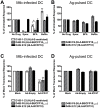
Figure 1
Figure 1. Mtb antigens are processed by the cytosolic pathway.
(A,B) Human monocyte-derived DC were treated with epoxomicin, BFA, or bafilomycin for one hour before infection with Mtb H37Rv-eGFP (A) or addition of CFP10 and CFP103–11 (B). After 15–16 hours in the presence of the inhibitor, DC were harvested, fixed, washed extensively, and used as APC in an IFN-γ ELISPOT assay where T cell clones are effectors. DC were added to an excess of T cells so that antigen was the limiting factor (see Materials and Methods). The mean number of spots produced by each clone was: D160 1-23 (228.4±39.3 to Mtb–infected DC, 19.6±6 to uninfected DC), D480 F6 (623.5±73.7 to Mtb–infected DC, 6.7±2.5 to uninfected DC), D454 E12 (453±52.4 to Mtb–infected DC, 13±4.4 to uninfected DC). Data have been normalized to the untreated control, and each bar reflects the mean±SEM of at least three experiments per clone (*p<0.05, **p<0.01 using two-tailed Student's t test compared to untreated controls, except where indicated). (C,D) DC were transduced with either empty vector or adenoviral ICP47 using Lipofectamine 2000. After 6–26 hours, DC were washed and infected with H37Rv-eGFP (C) or pulsed with antigen (D). Following overnight incubation, T cell clones were added and IFN-γ production was assessed by intracellular cytokine staining. The mean percentage of IFN-γ+ clones was: D160 1-23 (8.2±1.1 to Mtb–infected DC, 1.1±0.2 to uninfected DC), D480 F6 (46.4±3.9 to Mtb–infected DC, 1.5±0.4 to uninfected DC), D454 E12 (64±6 to Mtb–infected DC, 0.7±0.3 to uninfected DC). Each bar represents the mean±SEM of seven independent experiments.
Figure 2
Figure 2. Mtb proteins require retrotranslocation for presentation.
(A,B) DC were treated with exoA or cycloheximide for one hour prior to infection with H37Rv-eGFP (A) or addition of CFP10 and CFP103–11 (B). DC were harvested, fixed, and assessed for their ability to stimulate T cell clones by IFN-γ ELISPOT as described. Each bar reflects the mean±SEM of at least three experiments. ND, not done. (C) DC were treated with exoA, exoA/PJ34, or BSA/PJ34 for one hour prior to infection with vaccinia virus expressing eGFP. After 16–18 hours, DC were harvested and GFP expression analyzed by flow cytometry. Data are representative of three experiments. (D,E) DC treated with exoA, BSA, exoA/PJ34, or BSA/PJ34 for one hour were subsequently infected with H37Rv-eGFP (D) or pulsed with antigen (E) overnight, harvested, fixed, and assessed for their ability to stimulate T cell clones by IFN-γ ELISPOT. Data are representative of two experiments. (F) DC were treated with exoA, exoA/PJ34, or BSA/PJ34 for one hour prior to infection with vaccinia virus expressing HIV p24. After 16–18 hours, DC were harvested, fixed and used to stimulate the HIV p24306–316-specific CD8+ clone 16A7 in an IFN-y ELISPOT assay. Data are representative of two experiments.
Figure 3
Figure 3. The Mtb phagosome retains characteristics of an early endosome.
(A) Representative figure showing organelle distribution after percoll separation of homogenate from Mtb–infected DC. The plasma membrane was labeled with a PE-conjugated antibody to HLA-II prior to homogenization and fluorescence was detected by fluorometry. For detection of ER, fractions were assessed for the presence of TAP1 and PDI by western blot. An enzymatic assay for β-hexosaminidase was used for detection of lysosomes. Finally, fractions were examined for the presence of H37Rv-eGFP by flow cytometry and quantified using a reference latex bead population. For flow cytometric analysis of Mtb phagosomes, the final 2 ml (fractions 23–28) of the gradient were pelleted, fixed, permeabilized, and stained with antibodies of interest. (B) Magnetic bead and Mtb phagosomes were gated based on FSC/SSC (not shown) and then on LAMP-1/HLA-I (beads) or LAMP-1/GFP (Mtb). Arrows indicate the gated population. Analysis of phagosome maturation on one hour LAMP-1lo/−/HLA-I+ magnetic bead phagosomes (top panel), one hour LAMP-1+/HLA-Ilo/− magnetic bead phagosomes (second panel), overnight LAMP-1+ magnetic bead phagosomes (third panel), and overnight Mtb phagosomes (bottom panel). Plots include isotype staining (shaded histograms) as well as staining with the indicated antibody (red lines). The amplified HLA-I signal on the HLA-I-FITC gated events is due to the use of primary and secondary antibody combination, with which we routinely see up to a log shift in signal over conjugated primary. (C,D) Quantitative analysis of phagosomes over time. The percent positive number represents Overton cumulative histogram subtraction of the isotype control from the indicated stain. Each bar represents the mean±SEM of three experiments per timepoint.
Figure 4
Figure 4. The Mtb phagosome contains HLA-I loading accessory molecules.
(A) DC were pulsed with H37Rv-eGFP for 20 minutes, washed, and incubated for 40 minutes. Phagosomal fractions were prepared as in Figure 3 and stained with the indicated antibodies (top panel). Intact DC were fixed, permeabilized, and stained with the indicated antibodies (bottom panel). Data are representative of three experiments. (B) Quantitative analysis of Mtb phagosomes over time. The percent positive number represents Overton cumulative histogram subtraction of the isotype control from the indicated stain. Each bar represents the mean±SEM from three experiments per timepoint.
Figure 5
Figure 5. Mtb phagosomes contain minimal contamination.
(A) HLA-A2 or HLA-A2+ DC were infected with H37Rv-eGFP for 20 minutes, washed, and incubated for an additional 40 minutes. HLA-A2 DC were mixed with uninfected HLA-A2+ DC, homogenized, and homogenate separated using 27% percoll as described or pelleted without percoll separation. Phagosomes were stained with an antibody to HLA-A2. Shaded histograms represent isotype staining. Data are representative of three experiments. (B) HLA-A2 or HLA-A2+ LCL were fixed, permeabilized, and stained with an antibody to HLA-A2. (C,D) Mtb phagosomes (C) or intact DC (D) were analyzed for the presence of cis- and trans-golgi markers GM130 and golgin-97, respectively. Data in C are representative of three experiments each after a 40 minute or overnight chase. Data in D are representative of two experiments.
Figure 6
Figure 6. HLA-E:peptide complexes are present in phagosomal fractions.
(A,B) DC were pulsed with H37Rv-eGFP or CFP102–11 peptide and the homogenate was separated using 27% percoll as described. Each fraction was freeze-thawed and tested for its ability to stimulate D160 1-23 (A) or D160 1-1B (A&B) CD8+ T cell clones in the absence of additional APC. IFN-γ production was measured using ELISPOT. The mean±SEM of duplicate wells is presented. Data are representative of four experiments at various timepoints in A, and four experiments after a one hour peptide pulse in B. (C) Plasma membrane/ER fractions from Mtb–infected DC (HLA-B15+/HLA-B44) were incubated with HLA-I matched or mismatched CD8+ T cell clones and IFN-γ production was measured using ELISPOT. The mean±SEM of duplicate wells is presented and data are representative of three experiments done similarly. (D,E) Individual fractions were analyzed by flow cytometry to assess HLA-II-PE (plasma membrane) and H37Rv-eGFP fluorescence. Selected fractions are shown (D) including the peak plasma membrane fraction (#8) and phagosomal fractions (#24–27). Prior to flow cytometry, fractions were mixed with a reference latex bead population at a known concentration. Equal numbers of latex bead events were collected for all fractions and used to quantify the number of plasma membrane and phagosome particles (E) as described in Materials and Methods. (F) HLA-I was immunoprecipitated from plasma membrane or phagosome fractions using W6/32. After dilution of the plasma membrane sample to give similar levels of HC10 staining, the presence of HLA-I alleles was assessed using antibodies to pan-HLA-I (HC10), HLA-A2 (HCA2), and HLA-E (MEM-E/02). The numbers below blots indicate the relative intensity of the bands as described in Materials and Methods.
Figure 7
Figure 7. HLA-I, loading machinery, and HLA-I:peptide complexes are present in highly pure Mtb phagosomes.
(A) Phagosomes were isolated by percoll gradient or magnetic purification and prepared for electron microscopy as described in Materials and Methods. (B) Magnetic bead-isolated phagosomes were analyzed by flow cytometry to assess HLA-II-PE (plasma membrane) and H37Rv-eGFP fluorescence as described in Materials and Methods and Figure 6. The events shown represent a small proportion of the population of phagosomes isolated and the experiment is representative of three experiments. (C) DC were pulsed with magnetically-labeled H37Rv-eGFP for 20 minutes, washed, and incubated for 18 hr. After magnetic separation of Mtb phagosomes, flow organellometry was performed as described previously. Data are representative of three experiments. (D) Magnetically-isolated Mtb phagosomes were freeze-thawed and tested for their ability to stimulate D160 1-23 CD8+ T cell clones in the absence of additional APC. IFN-γ production was measured using ELISPOT. Data are representative of two experiments.

Similar articles

Cited by

References

    1. Corbett EL, Watt CJ, Walker N, Maher D, Williams BG, et al. The growing burden of tuberculosis: global trends and interactions with the HIV epidemic. Arch Intern Med. 2003;163:1009–1021. - PubMed
    1. Vergne I, Chua J, Singh SB, Deretic V. Cell biology of mycobacterium tuberculosis phagosome. Annu Rev Cell Dev Biol. 2004;20:367–394. - PubMed
    1. Flynn JL, Chan J. Immunology of tuberculosis. Annu Rev Immunol. 2001;19:93–129. - PubMed
    1. Grotzke JE, Lewinsohn DM. Role of CD8+ T lymphocytes in control of Mycobacterium tuberculosis infection. Microbes Infect. 2005;7:776–788. - PubMed
    1. Shen L, Rock KL. Priming of T cells by exogenous antigen cross-presented on MHC class I molecules. Curr Opin Immunol. 2006;18:85–91. - PubMed

Publication types

MeSH terms

Substances