. 2009:7161:nihpa96839.
doi: 10.1117/12.808314.

Human bladder cancer diagnosis using Multiphoton microscopy

Affiliations

Human bladder cancer diagnosis using Multiphoton microscopy

Sushmita Mukherjee et al. Proc SPIE Int Soc Opt Eng. 2009.

Abstract

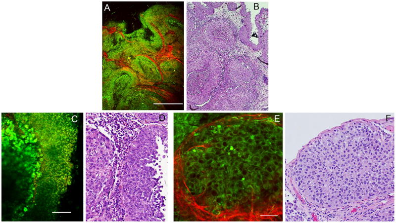
At the time of diagnosis, approximately 75% of bladder cancers are non-muscle invasive. Appropriate diagnosis and surgical resection at this stage improves prognosis dramatically. However, these lesions, being small and/or flat, are often missed by conventional white-light cystoscopes. Furthermore, it is difficult to assess the surgical margin for negativity using conventional cystoscopes. Resultantly, the recurrence rates in patients with early bladder cancer are very high. This is currently addressed by repeat cystoscopies and biopsies, which can last throughout the life of a patient, increasing cost and patient morbidity. Multiphoton endoscopes offer a potential solution, allowing real time, non-invasive biopsies of the human bladder, as well as an up-close assessment of the resection margin. While miniaturization of the Multiphoton microscope into an endoscopic format is currently in progress, we present results here indicating that Multiphoton imaging (using a bench-top Multiphoton microscope) can indeed identify cancers in fresh, unfixed human bladder biopsies. Multiphoton images are acquired in two channels: (1) broadband autofluorescence from cells, and (2) second harmonic generation (SHG), mostly by tissue collagen. These images are then compared with gold standard hematoxylin/eosin (H&E) stained histopathology slides from the same specimen. Based on a "training set" and a very small "blinded set" of samples, we have found excellent correlation between the Multiphoton and histopathological diagnoses. A larger blinded analysis by two independent uropathologists is currently in progress. We expect that the conclusion of this phase will provide us with diagnostic accuracy estimates, as well as the degree of inter-observer heterogeneity.


Figures

Figure 1
Figure 1
Multiphoton images and Hematoxylin/eosin (H&E) stained histopathology slides showing comparable architecture in biopsy with normal histology. A: Multiphoton image at low magnification, acquired with a 4X (NA 0.28) dry objective. Color-coding: Green = broadband autofluorescence; Red: Second Harmonic Generation (SHG) signal. Scale bar = 500 μm. B: Image acquired from a corresponding area of an H&E stained slide prepared from the same specimen. C: Multiphoton image at high magnification, acquired with a 20X (NA 0.95) water immersion objective. Color-coding: Green = broadband autofluorescence; Red: Second Harmonic Generation (SHG) signal. Scale bar = 100 μm. D: Image acquired from a corresponding area of an H&E stained slide prepared from the same specimen.
Figure 2
Figure 2
Multiphoton images and Hematoxylin/eosin (H&E) stained histopathology slides showing comparable architecture in a low-grade papillary lesion. A: Multiphoton image at low magnification, acquired with a 4X (NA 0.28) dry objective. Color-coding: Green = broadband autofluorescence; Red: Second Harmonic Generation (SHG) signal. Scale bar = 500 μm. B: Image acquired from a corresponding area of an H&E stained slide prepared from the same specimen. C: Multiphoton image at high magnification, acquired with a 20X (NA 0.95) water immersion objective. Color-coding: Green = broadband autofluorescence; Red: Second Harmonic Generation (SHG) signal. Scale bar = 100 μm. D: Image acquired from a corresponding area of an H&E stained slide prepared from the same specimen. E: Multiphoton image at high magnification at a second location, acquired with a 20X (NA 0.95) water immersion objective. Color-coding: Green = broadband autofluorescence; Red: Second Harmonic Generation (SHG) signal. Scale bar = 100 μm. F: Image acquired from a corresponding area of an H&E stained slide prepared from the same specimen.
Figure 3
Figure 3
Multiphoton images and Hematoxylin/eosin (H&E) stained histopathology slides showing comparable architecture in a high-grade papillary lesion. A: Multiphoton image at low magnification, acquired with a 4X (NA 0.28) dry objective. Color-coding: Green = broadband autofluorescence; Red: Second Harmonic Generation (SHG) signal. Scale bar = 500 μm. B: Image acquired from a corresponding area of an H&E stained slide prepared from the same specimen. C: Multiphoton image at high magnification, acquired with a 20X (NA 0.95) water immersion objective. Color-coding: Green = broadband autofluorescence; Red: Second Harmonic Generation (SHG) signal. Scale bar = 100 μm. D: Image acquired from a corresponding area of an H&E stained slide prepared from the same specimen. E: Multiphoton image at high magnification at a second location, acquired with a 20X (NA 0.95) water immersion objective. Color-coding: Green = broadband autofluorescence; Red: Second Harmonic Generation (SHG) signal. Scale bar = 50 μm (a digital zoom of 2 was used). F: Image acquired from a corresponding area of an H&E stained slide prepared from the same specimen.

Similar articles

Cited by

References

    1. “American Cancer Society: Cancer Facts and Figures”. (1995).

    1. Nieder AM, Soloway MS. Eliminate the term “superficial” bladder cancer. Journal of Urology. 2006;175(2):417–8. - PubMed
    1. Lee CSD, Yoon CY, Witjes JA. The past, present and future of cystoscopy: the fusion of cystoscopy and novel imaging technology. BJU International. 2008;102:1228–33. - PubMed
    1. Kwast TH. Urine markers for bladder cancer surveillance: a systematic review. European Urology. 2005;47:736–48. - PubMed
    1. Botteman MF, et al. The health economics of bladder cancer: a comprehensive review of the published literature. Pharmacoeconomics. 2003;21(18):1315–30. - PubMed

LinkOut - more resources