

Expression and location of alpha-fetoprotein during rat colon development
- PMID: 19360917
- PMCID: PMC2668779
- DOI: 10.3748/wjg.15.1738
Expression and location of alpha-fetoprotein during rat colon development
Abstract
Aim: To investigate the expression of alpha-fetoprotein (AFP), a cancer-associated fetal glycoprotein, and its involvement during rat colon development.
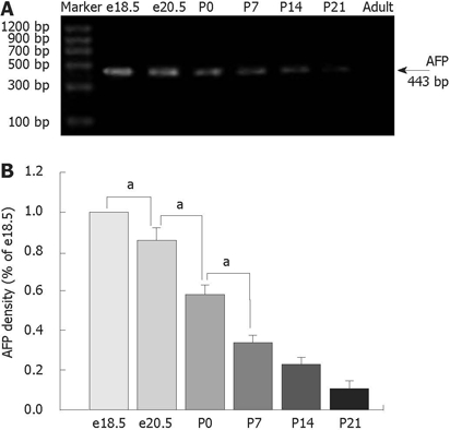
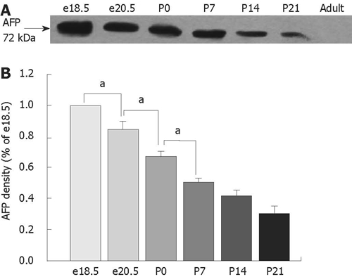
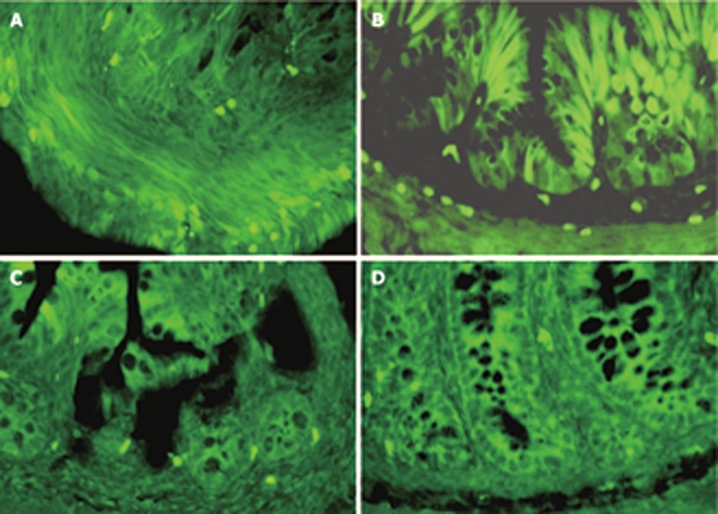
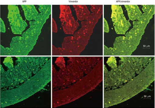
Methods: Colons from Sprague-Dawley rat fetuses, young and adult (8 wk old) animals were used in this study. Expression levels of AFP in colons of different development stage were detected by reverse-transcriptase PCR (RT-PCR) and Western blotting. To identify the cell location of AFP in the developing rat colons, double-immunofluorescent staining was performed using antibodies to specific cell markers and AFP, respectively.
Results: The highest levels of AFP mRNA were detected in colons of rats at embryonic day 18.5 (e18.5). Compared to e18.5 d, the AFP expression was significantly decreased during rat development [85% for e20.5, P < 0.05, 58% for postnatal day 0.5 (P0.5), P < 0.05, 37% for P7, P < 0.05, 24% for P14, P < 0.05, and 11% for P21, P < 0.05] and undetected in adult rats. Only the 72-kDa isoform of AFP was detected by Western blotting, the expression pattern was similar to AFP mRNA and conformed to the results of mRNA expression. The AFP positive staining was identical to different distribution patterns in fetuses, young and adult animals and positive staining for both AFP and vimentin was overlapped in mesenchymal cells at each stage tested.
Conclusion: This study has for the first time demonstrated that AFP is localized in the mesenchyme of rat colon from the embryo to the weaning stage by immunofluorescence and presents 72-kDa isoform in the developing rat colons by Western blotting. The dynamic expression of AFP in the various developmental stages of the colon indicates that AFP might be involved in many aspects of colon development.
Figures
Similar articles
-
Expression and localization of Wolfram syndrome 1 gene in the developing rat pancreas.World J Gastroenterol. 2009 Nov 21;15(43):5425-31. doi: 10.3748/wjg.15.5425. World J Gastroenterol. 2009. PMID: 19916172 Free PMC article.
-
α-fetoprotein involvement during glucocorticoid-induced precocious maturation in rat colon.World J Gastroenterol. 2011 Jun 28;17(24):2933-40. doi: 10.3748/wjg.v17.i24.2933. World J Gastroenterol. 2011. PMID: 21734804 Free PMC article.
-
Alpha-fetoprotein is dynamically expressed in rat pancreas during development.Dev Growth Differ. 2007 Oct;49(8):669-81. doi: 10.1111/j.1440-169X.2007.00961.x. Dev Growth Differ. 2007. PMID: 17880577
-
Expression and localization of paxillin in rat pancreas during development.World J Gastroenterol. 2011 Oct 28;17(40):4479-87. doi: 10.3748/wjg.v17.i40.4479. World J Gastroenterol. 2011. PMID: 22110278 Free PMC article.
-
[Several transcription factors participate in the functioning of the alpha-fetoprotein gene promoter].Bull Cancer. 1995 Jul;82(7):541-50. Bull Cancer. 1995. PMID: 7549116 Review. French.
Cited by
-
Expression and localization of Wolfram syndrome 1 gene in the developing rat pancreas.World J Gastroenterol. 2009 Nov 21;15(43):5425-31. doi: 10.3748/wjg.15.5425. World J Gastroenterol. 2009. PMID: 19916172 Free PMC article.
-
α-fetoprotein involvement during glucocorticoid-induced precocious maturation in rat colon.World J Gastroenterol. 2011 Jun 28;17(24):2933-40. doi: 10.3748/wjg.v17.i24.2933. World J Gastroenterol. 2011. PMID: 21734804 Free PMC article.
References
-
- Liu JP, Baker J, Perkins AS, Robertson EJ, Efstratiadis A. Mice carrying null mutations of the genes encoding insulin-like growth factor I (Igf-1) and type 1 IGF receptor (Igf1r) Cell. 1993;75:59–72. - PubMed
-
- Luetteke NC, Qiu TH, Peiffer RL, Oliver P, Smithies O, Lee DC. TGF alpha deficiency results in hair follicle and eye abnormalities in targeted and waved-1 mice. Cell. 1993;73:263–278. - PubMed
-
- Weinstein DC, Ruiz i Altaba A, Chen WS, Hoodless P, Prezioso VR, Jessell TM, Darnell JE Jr. The winged-helix transcription factor HNF-3 beta is required for notochord development in the mouse embryo. Cell. 1994;78:575–588. - PubMed
-
- Schmidt C, Bladt F, Goedecke S, Brinkmann V, Zschiesche W, Sharpe M, Gherardi E, Birchmeier C. Scatter factor/hepatocyte growth factor is essential for liver development. Nature. 1995;373:699–702. - PubMed
MeSH terms
Substances
LinkOut - more resources
Full Text Sources
Research Materials
