

Design, synthesis, and preclinical evaluation of prostate-specific membrane antigen targeted (99m)Tc-radioimaging agents
- PMID: 19361232
- PMCID: PMC9190123
- DOI: 10.1021/mp9000712
Design, synthesis, and preclinical evaluation of prostate-specific membrane antigen targeted (99m)Tc-radioimaging agents
Abstract
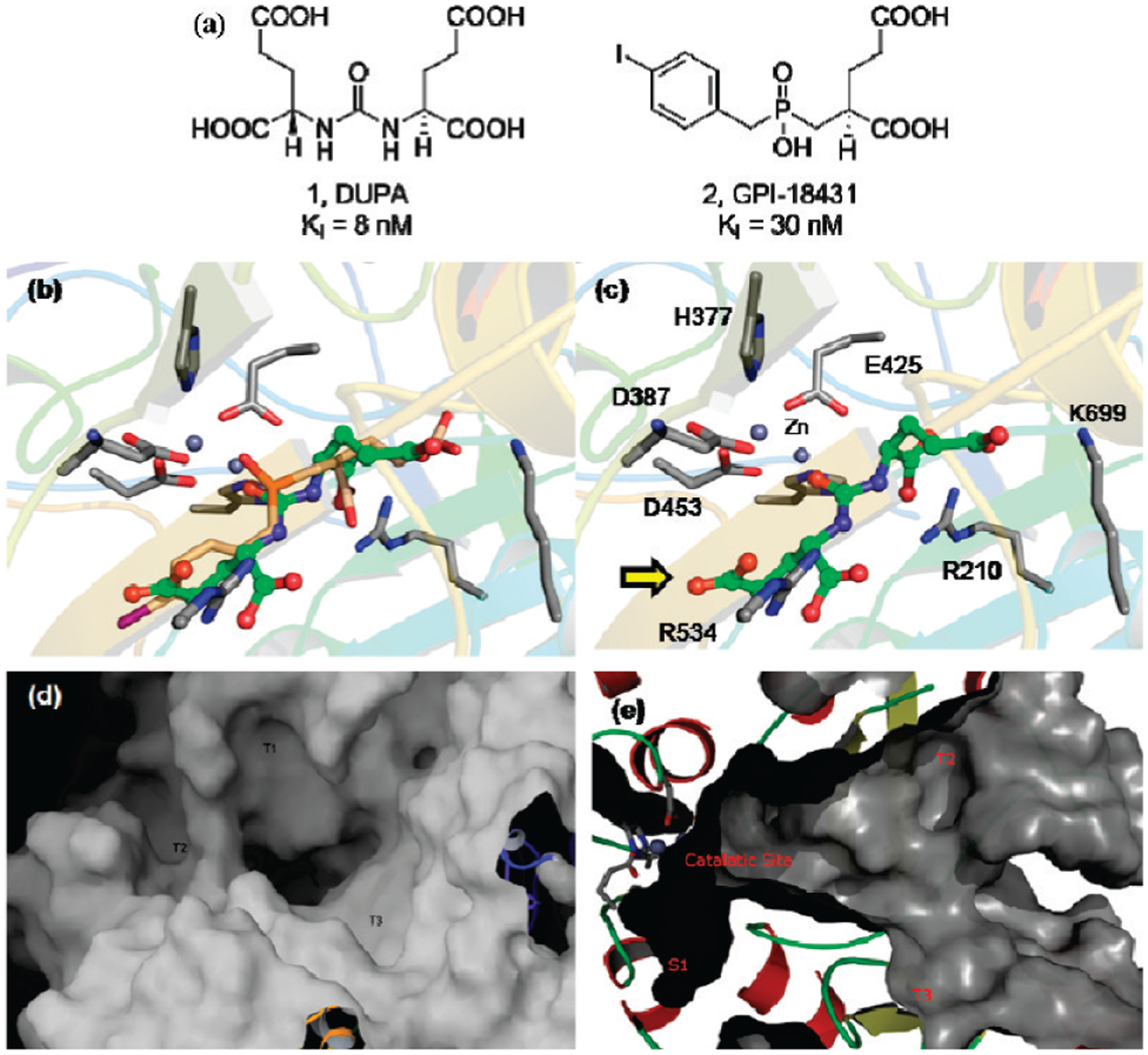
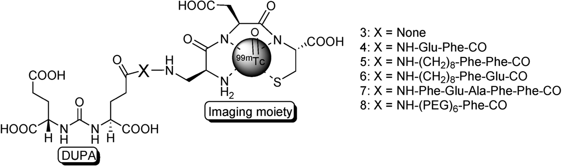
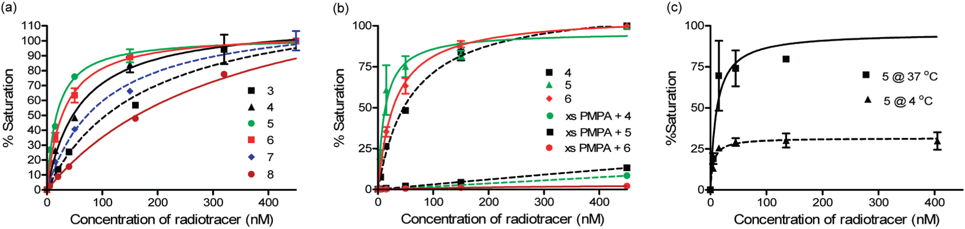
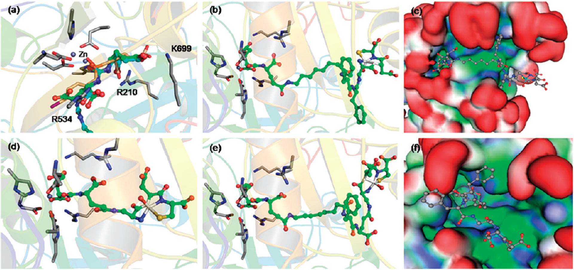
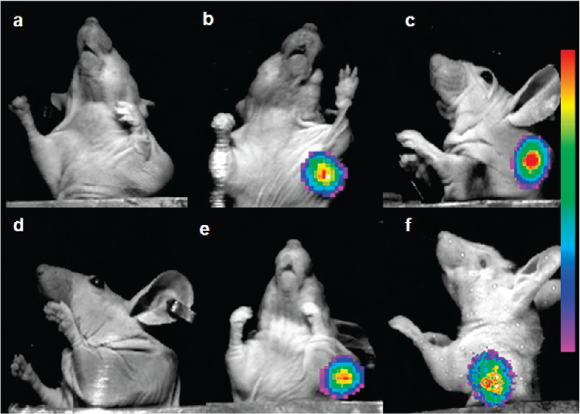
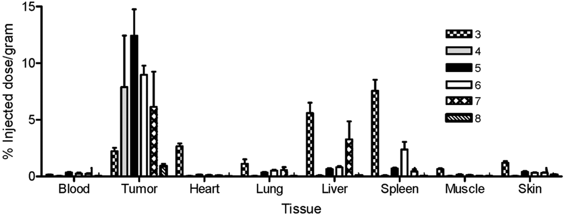
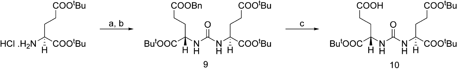
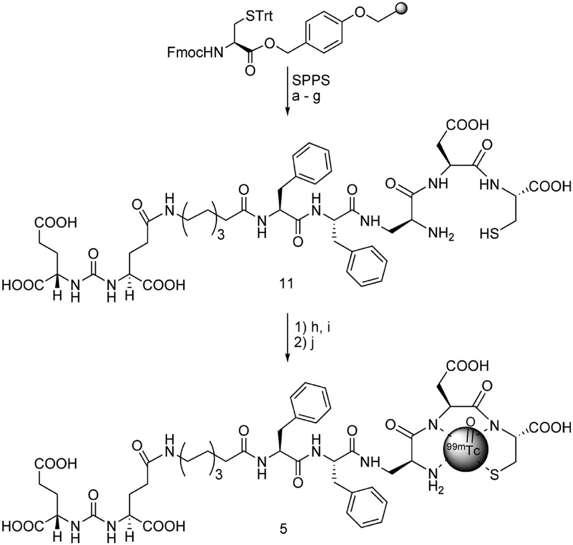
The high mortality and financial burden associated with prostate cancer can be partly attributed to a lack of sensitive screening methods for detection and staging of the disease. Guided by in silico docking studies using the crystal structure of PSMA, we designed and synthesized a series of PSMA-targeted (99m)Tc-chelate complexes for imaging PSMA-expressing human prostate cancer cells (LNCaP cell line). Of the six targeted radioimaging agents synthesized, three were found to bind LNCaP cells with low nanomolar affinity. Moreover, the same three PSMA-targeted imaging agents were shown to localize primarily to LNCaP tumor xenografts in nu/nu mice, with an average of 9.8 +/- 2.4% injected dose/g tissue accumulating in the tumor and only 0.11% injected dose/g tissue retained in the muscle at 4 h postinjection. Collectively, these high affinity, PSMA-specific radioimaging agents demonstrate significant potential for use in localizing prostate cancer masses, monitoring response to therapy, detecting prostate cancer recurrence following surgery, and selecting patients for subsequent PSMA-targeted chemotherapy.
Figures
Similar articles
-
Prostate-specific membrane antigen targeted imaging and therapy of prostate cancer using a PSMA inhibitor as a homing ligand.Mol Pharm. 2009 May-Jun;6(3):780-9. doi: 10.1021/mp900069d. Mol Pharm. 2009. PMID: 19361233
-
Preparation and Biological Evaluation of [99mTc]Tc-CNGU as a PSMA-Targeted Radiotracer for the Imaging of Prostate Cancer.Molecules. 2020 Nov 26;25(23):5548. doi: 10.3390/molecules25235548. Molecules. 2020. PMID: 33256058 Free PMC article.
-
Novel technetium-99m-labeled bivalent PSMA-targeting probe based on hydroxamamide chelate for diagnosis of prostate cancer.Ann Nucl Med. 2024 Oct;38(10):847-851. doi: 10.1007/s12149-024-01959-9. Epub 2024 Jul 8. Ann Nucl Med. 2024. PMID: 38976087
-
Inhibitors of prostate-specific membrane antigen in the diagnosis and therapy of metastatic prostate cancer - a review of patent literature.Expert Opin Ther Pat. 2021 Jun;31(6):525-547. doi: 10.1080/13543776.2021.1878145. Epub 2021 Apr 16. Expert Opin Ther Pat. 2021. PMID: 33459068 Review.
-
The Rise of PSMA Ligands for Diagnosis and Therapy of Prostate Cancer.J Nucl Med. 2016 Oct;57(Suppl 3):79S-89S. doi: 10.2967/jnumed.115.170720. J Nucl Med. 2016. PMID: 27694178 Review.
Cited by
-
Engineering a prostate-specific membrane antigen-activated tumor endothelial cell prodrug for cancer therapy.Sci Transl Med. 2012 Jun 27;4(140):140ra86. doi: 10.1126/scitranslmed.3003886. Sci Transl Med. 2012. PMID: 22745436 Free PMC article. Clinical Trial.
-
[(68)Ga-PSMA as a new tracer for evaluation of prostate cancer: comparison between PET-CT and PET-MRI in biochemical recurrence].Radiologe. 2015 Feb;55(2):89-91. doi: 10.1007/s00117-014-2792-6. Radiologe. 2015. PMID: 25583413 German. No abstract available.
-
Preclinical Comparative Study of (68)Ga-Labeled DOTA, NOTA, and HBED-CC Chelated Radiotracers for Targeting PSMA.Bioconjug Chem. 2016 Jun 15;27(6):1447-55. doi: 10.1021/acs.bioconjchem.5b00679. Epub 2016 May 9. Bioconjug Chem. 2016. PMID: 27076393 Free PMC article.
-
A small molecule drug conjugate (SMDC) of DUPA and a duocarmycin built on the solid phase.Medchemcomm. 2019 Nov 27;10(12):2170-2174. doi: 10.1039/c9md00279k. eCollection 2019 Dec 1. Medchemcomm. 2019. PMID: 32879717 Free PMC article.
-
[99mTc]Tc-PSMA-T4-Novel SPECT Tracer for Metastatic PCa: From Bench to Clinic.Molecules. 2022 Oct 25;27(21):7216. doi: 10.3390/molecules27217216. Molecules. 2022. PMID: 36364046 Free PMC article.
References
-
- Jemal A; Siegel R; Ward E Cancer statistics 2008. Ca–Cancer J. Clin 2008, 58, 71–96. - PubMed
-
- Zeller JL Grading of prostate cancer. JAMA, J. Am. Med. Assoc 2007, 298, 1596. - PubMed
-
- Chodak MD; Keller P; Schoenberg HW Assessment of screening for prostate cancer using the digital rectal examination. J. Urol 1989, 141, 1136–1138. - PubMed
-
- Essink-Bot M; de Koning HJ; Nijs HGJ; Kirkels WJ; van der Mass PJ; Schroder FH Short term effects of population-based screening for prostate cancer on health-related quality of life. J. Natl. Cancer Inst 1998, 90, 925–931. - PubMed
-
- Linn MM; Ball RA; Maradiegue A Prostate-specific antigen screening: Friend or foe? Urol. Nurs 2007, 27, 481–489. - PubMed
Publication types
MeSH terms
Substances
Grants and funding
LinkOut - more resources
Full Text Sources
Other Literature Sources
Medical
Research Materials
Miscellaneous
