

Task-free presurgical mapping using functional magnetic resonance imaging intrinsic activity
- PMID: 19361264
- PMCID: PMC3686552
- DOI: 10.3171/2008.10.JNS08846
Task-free presurgical mapping using functional magnetic resonance imaging intrinsic activity
Abstract
Object: Low-frequency components of the spontaneous functional MR imaging signal provide information about the intrinsic functional and anatomical organization of the brain. The ability to use such methods in individual patients may provide a powerful tool for presurgical planning. The authors explore the feasibility of presurgical motor function mapping in which a task-free paradigm is used.
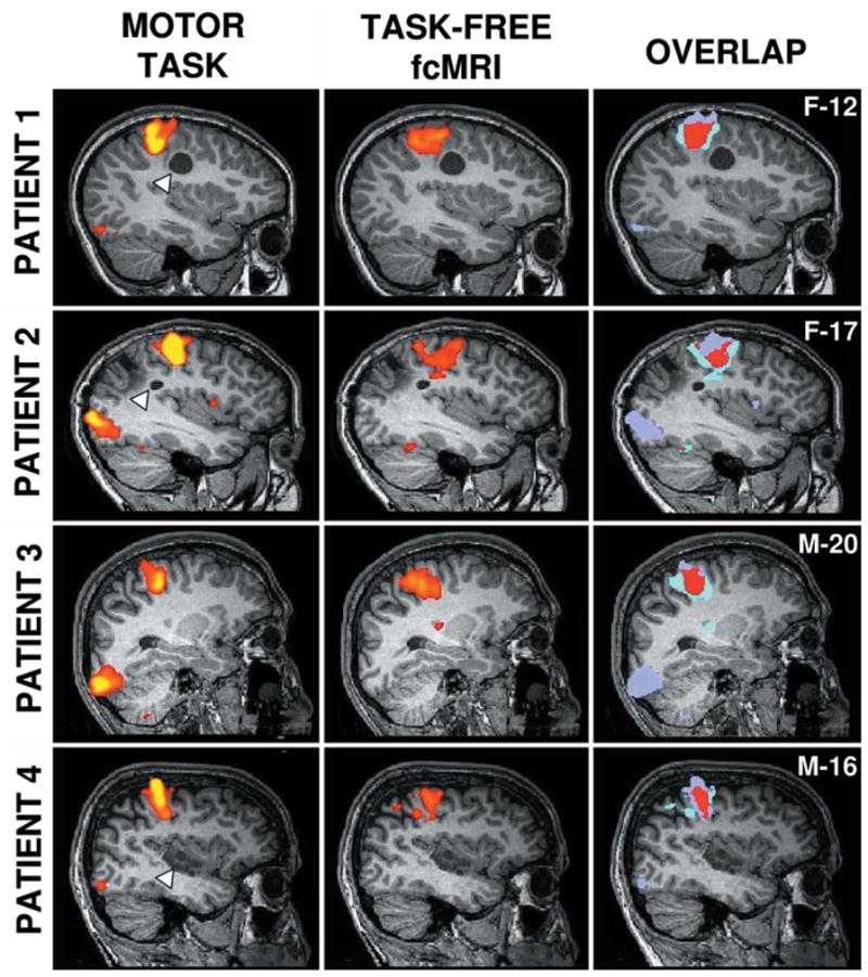
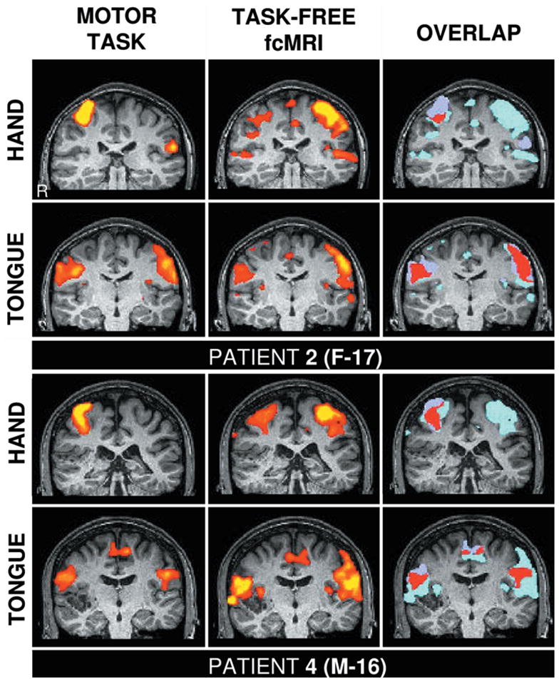
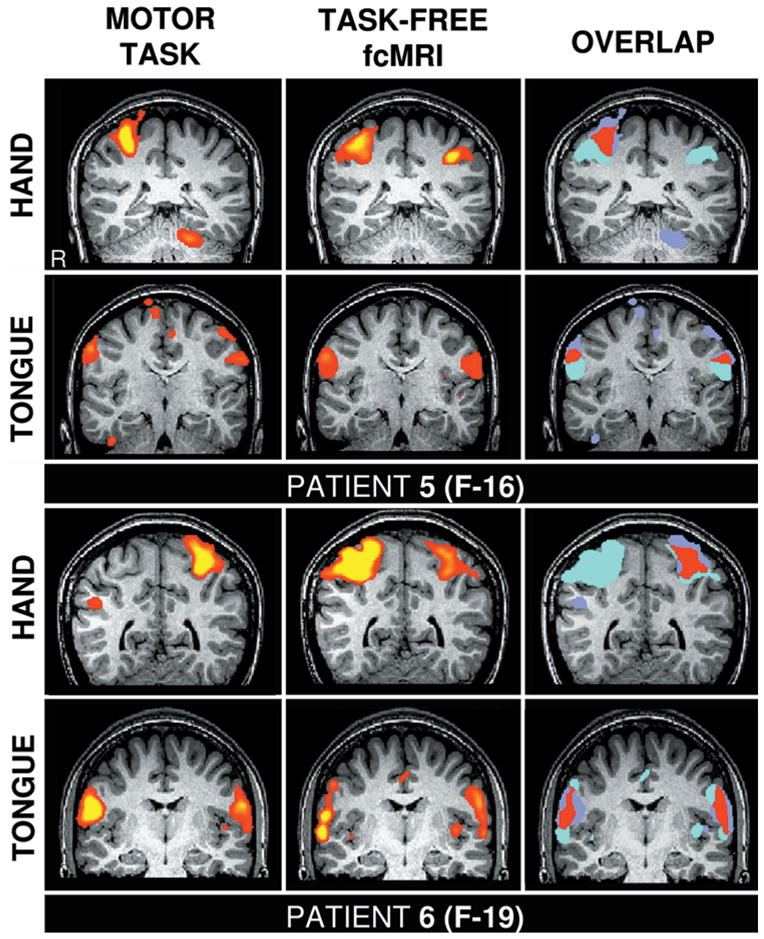
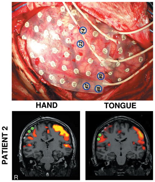
Methods: Six surgical candidates with tumors or epileptic foci near the motor cortex participated in this study. The investigators directly compared task-elicited activation of the motor system to activation obtained from intrinsic activity correlations. The motor network within the unhealthy hemisphere was identified based on intrinsic activity correlations, allowing distortions of functional anatomy caused by the tumor and epilepsy to be directly visualized. The precision of the motor function mapping was further explored in 1 participant by using direct cortical stimulation.
Results: The motor regions localized based on the spontaneous activity correlations were quite similar to the regions defined by actual movement tasks and cortical stimulation. Using intrinsic activity correlations, it was possible to map the motor cortex in presurgical patients.
Conclusions: This task-free paradigm may provide a powerful approach to map functional anatomy in patients without task compliance and allow multiple brain systems to be determined in a single scanning session.
Figures
References
-
- Abou–Khalil B, Schlaggar BL. Is it time to replace the Wada test? Neurology. 2002;59:160–161. - PubMed
-
- Berger MS, Cohen WA, Ojemann GA. Correlation of motorcortex brain mapping data with magnetic resonance imaging. J Neurosurg. 1990;72:383–387. - PubMed
-
- Biswal B, Yetkin FZ, Haughton VM, Hyde JS. Functional connectivity in the motor cortex of resting human brain using echo-planar MRI. Magn Reson Med. 1995;34:537–554. - PubMed
-
- Bittar RG, Olivier A, Sadikot AF, Andermann F, Pike GB, Reutens DC. Presurgical motor and somatosensory cortex mapping with functional magnetic resonance imaging and positron emission tomography. J Neurosurg. 1999;91:915–921. - PubMed
-
- Boroojerdi B. Localization of the motor hand area using transcranial magnetic stimulation and functional magnetic resonance imaging. Clin Neurophysiol. 1999;110:699–704. - PubMed
Publication types
MeSH terms
Grants and funding
LinkOut - more resources
Full Text Sources
Medical
