Review
. 2009 Sep;11(9):2245-63.
doi: 10.1089/ars.2009.2611.

Redox regulation of tumor necrosis factor signaling

Affiliations
Review

Redox regulation of tumor necrosis factor signaling

Derick Han et al. Antioxid Redox Signal. 2009 Sep.

Abstract

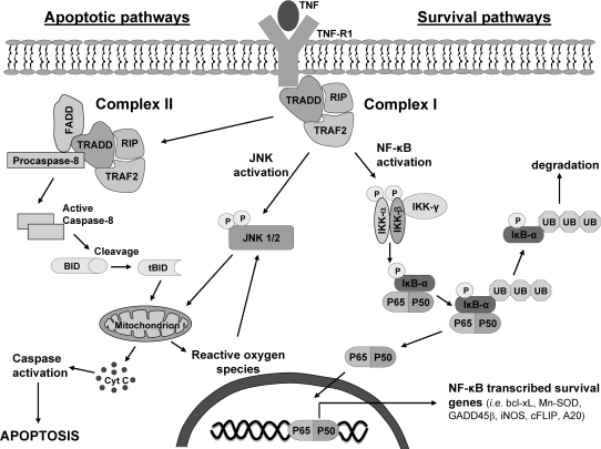
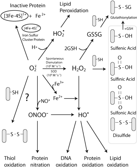
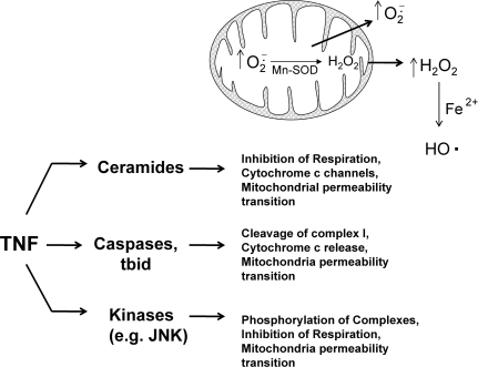
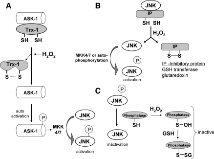
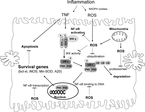
Tumor necrosis factor-alpha (TNF) is a key cytokine that has been shown to play important physiologic (e.g., inflammation) and pathophysiologic (e.g., various liver pathologies) roles. In liver and other tissues, TNF treatment results in the simultaneous activation of an apoptotic pathway (i.e., TRADD, RIP, JNK) and a survival pathway mediated by NF-kappaB transcription of survival genes (i.e., GADD45beta, Mn-SOD, cFLIP). The cellular response (e.g., proliferation versus apoptosis) to TNF is determined by the balance between the apoptotic signaling pathway and the NF-kappaB survival pathway stimulated by TNF. Reactive oxygen species (ROS) are important modulators of signaling pathways and can regulate both apoptotic signaling and NF-kappaB transcription triggered by TNF. ROS are important in mediating the sustained activation of JNK, to help mediate apoptosis after TNF treatment. In some cells, ROS are second messengers that mediate apoptosis after TNF stimulation. Conversely, ROS can cause redox modifications that inhibit NF-kappaB activation, which can lead to cell death triggered by TNF. Consequently, the redox status of cells can determine the biologic response that TNF will induce in cells. In many liver pathologies, ROS generated extrinsically (e.g., inflammation) or intrinsically (i.e., drugs, toxins) may act in concert with TNF to promote hepatocyte death and liver injury through redox inhibition of NF-kappaB.


Figures

FIG. 1.
FIG. 1.
Activation of apoptotic and survival pathways by TNF. The pleiotropic biologic effects of TNF can be attributed to its ability to activate apoptotic and survival pathways simultaneously. The apoptotic pathway is initialized by the formation of complex I, which is eventually internalized to form complex II. Complex II activates caspase-8, which cleaves Bid to tBid, which translocates to mitochondria and permeabilizes the mitochondrial outer membrane to allow cytochrome c release and possibly to increase mitochondrial ROS generation. The newly released cytochrome c can further activate other caspases, which can target mitochondria, leading to a positive-feedback loop, resulting in extensive caspase activation and ROS generation and ultimately apoptosis. TRAF-2 in complex I also activates the MAP kinase cascade [ASK-1, MKK4/7 (not shown)], leading to the activation of JNK, important in mediating apoptosis in some cells. After activation, JNK translocates to mitochondria and promotes cytochrome c release, mitochondrial permeability transition, and possibly increased ROS generation. Because JNK can be activated by ROS, a positive-feedback loop may ensue in which JNK translocation to mitochondria causes increased ROS generation, which activates more JNK molecules. The survival pathway is mediated by NF-κB transcription of survival genes that block many proteins involved in apoptosis. Complex I will activate IKK, which phosphorylates IκB-α, promoting its ubiquitination and degradation. The degradation of IκB-α releases NF-κB, allowing NF-κB to translocate to the nucleus and promote transcription of survival genes. Consequently, the transcription of NF-κB–regulated proteins, not apoptosis, is the major response to TNF in hepatocytes and other primary cells, under normal conditions.
FIG. 2.
FIG. 2.
Redox changes mediated by formula image and H2O2 in cells. formula image can be generated by many sources in cells, including NADPH oxidase and the mitochondrial respiratory chain. formula image can mediate many reactions that modulate cellular redox status and signaling pathways: (a) formula image can oxidize proteins with iron-sulfur clusters such as aconitase and other metal groups to modulate activity and function; (b) formula image can reduce transition metals (e.g., Fe2+ to Fe3+) important in catalyzing the formation of hydroxyl radical (the most reactive and damaging radical) from H2O2; (c) when nitric oxide is present, formula image will react with nitric oxide to generate ONOO, a potent oxidant; (d) formula image reactivity increases after protonated formula image (perhydroxyl radical), which can initiate free radical chain reactions such as lipid peroxidation; and (e) formula image can oxidize thiols to disulfides, but whether the reaction rates are physiologically relevant is debated. formula image will, spontaneously or through actions of SOD, dismutate into H2O2. H2O2 can readily oxidize cysteines in proteins to disulfides and sulfenic, sulfinic, or sulfonic acids, which can affect protein activity. It is believed that H2O2 modifies signaling pathways through posttranslational modification of cysteine and other amino acids (e.g., methionine) on proteins.
FIG. 3.
FIG. 3.
Possible pathways activated by TNF that mediate ROS generation from mitochondria. During apoptosis stimulated by TNF, mitochondrial respiration is inhibited, and ROS generation increases in most cells. Several potential signaling pathways are activated during TNF signaling that can potentially affect mitochondrial respiration and ROS generation, including (a) ceramides, signaling lipids that regulate mitochondria function; (b) kinases such JNK, which translocate to mitochondria and phosphorylate mitochondrial proteins; and (c) caspases and proapoptotic bcl-2 family members, which can cleave respiratory complexes and inhibit mitochondrial function. All these pathways can inhibit respiration, potentially to cause increased ROS generation from mitochondria, a major source of both formula image and H2O2 in cells.
FIG. 4.
FIG. 4.
Redox regulation of JNK. JNK is activated by ROS in most cells through redox regulation of proteins involved in its regulation. The sustained JNK activity by ROS may be due to (A) Trx1 oxidation causing the disassociation from ASK-1. JNK is primarily phosphorylated by MKK (a MAPK kinase), which in turn is phosphorylated by ASK-1 (a MAPK kinase kinase) in liver. ASK-1 is inhibited in cytoplasm by an association with Trx-1, which contains critical redox-sensitive thiols. The oxidation of these critical thiols on Trx-1 by H2O2 or other oxidants will cause Trx-1 to disassociate from ASK-1, which subsequently self-activates. Once activated, ASK-1 will phosphorylate MKK, which then activates JNK. (B) Oxidation of JNK inhibitory proteins. JNK is inhibited through association with redox-regulated proteins such as glutaredoxin and glutathione S-transferase, which contain critical cysteines. ROS can oxidize these inhibitory proteins, liberating JNK, which subsequently becomes activated by MKK or through autophosphorylation. (C) Redox inhibition of MAP kinase phosphatase. MAP kinase phosphatase, important in dephosphorylating and inactivating JNK, contains critical thiols that regulate its activity. Sustained JNK activity by ROS can be a result of redox inhibition of phosphatase through oxidation of its key thiols (i.e., sulfenic acid and disulfide formation, such as glutathionylation).
FIG. 5.
FIG. 5.
Potential sites in NF-κB signaling that are redox regulated. Many sites involved in NF-κB signaling are redox regulated: (a) IKK, important in phosphorylation of IκB-α (necessary for its degradation) can be inhibited by redox changes (cysteine-179); (b) proteins involved in ubiquitination of IκB-α and proteasome activity, necessary for its degradation, have been shown to be inhibited by ROS; (c) NF-κB has critical thiols (cysteine-62 on p50) important in binding to DNA; and (d) NF-κB transcription of survival genes also was shown to be inhibited by ROS and GSH depletion. Inflammation, which produces both TNF and ROS, may trigger apoptosis in hepatocytes and other primary cells, through redox inhibition of NF-κB signaling.

References

    1. Adler V. Yin Z. Fuchs SY. Benezra M. Rosario L. Tew KD. Pincus MR. Sardana M. Henderson CJ. Wolf CR. Davis RJ. Ronai Z. Regulation of JNK signaling by GSTp. EMBO J. 1999;18:1321–1334. - PMC - PubMed
    1. Antosiewicz J. Ziolkowski W. Kaczor JJ. Herman-Antosiewicz A. Tumor necrosis factor-alpha-induced reactive oxygen species formation is mediated by JNK1-dependent ferritin degradation and elevation of labile iron pool. Free Radic Biol Med. 2007;43:265–270. - PubMed
    1. Antras-Ferry J. Maheo K. Morel F. Guillouzo A. Cillard P. Cillard J. Dexamethasone differently modulates TNF-alpha- and IL-1beta-induced transcription of the hepatic Mn-superoxide dismutase gene. FEBS Lett. 1997;403:100–104. - PubMed
    1. Antunes F. Han D. Cadenas E. Relative contributions of heart mitochondria glutathione peroxidase and catalase to H(2)O(2) detoxification in in vivo conditions. Free Radic Biol Med. 2002;33:1260–1267. - PubMed
    1. Antunes F. Salvador A. Marinho HS. Alves R. Pinto RE. Lipid peroxidation in mitochondrial inner membranes, I: an integrative kinetic model. Free Radic Biol Med. 1996;21:917–943. - PubMed

Publication types

Substances