. 2009 Aug;23(6):851-60.
doi: 10.1016/j.bbi.2009.04.001. Epub 2009 Apr 8.

Stress exacerbates neuropathic pain via glucocorticoid and NMDA receptor activation

Affiliations

Stress exacerbates neuropathic pain via glucocorticoid and NMDA receptor activation

Jessica K Alexander et al. Brain Behav Immun. 2009 Aug.

Abstract

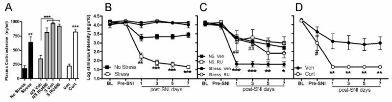
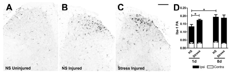
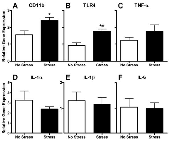
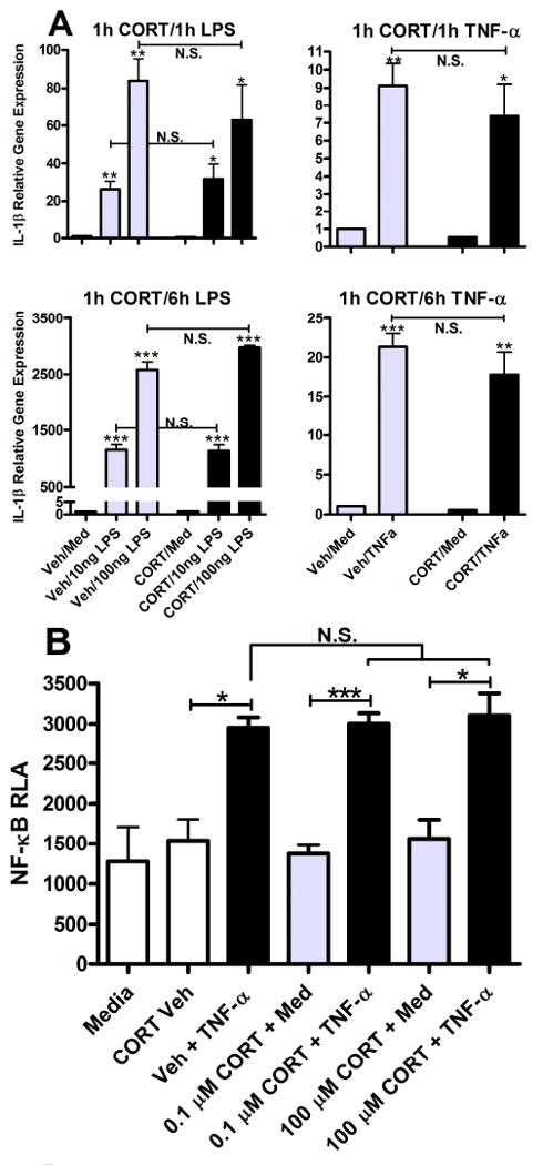
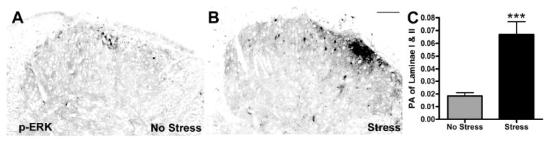
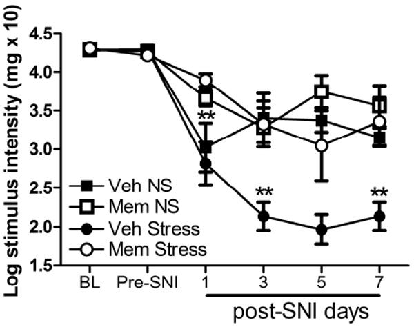
There is growing recognition that psychological stress influences pain. Hormones that comprise the physiological response to stress (e.g., corticosterone; CORT) may interact with effectors of neuropathic pain. To test this hypothesis, mice received a spared nerve injury (SNI) after exposure to 60 min restraint stress. In stressed mice, allodynia was consistently increased. The mechanism(s) underlying the exacerbated pain response involves CORT acting via glucocorticoid receptors (GRs); RU486, a GR antagonist, prevented the stress-induced increase in allodynia whereas exogenous administration of CORT to non-stressed mice reproduced the allodynic response caused by stress. Since nerve injury-induced microglial activation has been implicated in the onset and propagation of neuropathic pain, we evaluated cellular and molecular indices of microglial activation in the context of stress. Activation of dorsal horn microglia was accelerated by stress; however, this effect was transient and was not associated with the onset or maintenance of a pro-inflammatory phenotype. Stress-enhanced allodynia was associated with increased dorsal horn extracellular signal-regulated kinase phosphorylation (pERK). ERK activation could indicate a stress-mediated increase in glutamatergic signaling, therefore mice were treated prior to SNI and stress with memantine, an N-methyl-D-aspartate receptor (NMDAR) antagonist. Memantine prevented stress-induced enhancement of allodynia after SNI. These data suggest that the hormonal responses elicited by stress exacerbate neuropathic pain through enhanced central sensitization. Moreover, drugs that inhibit glucocorticoids (GCs) and/or NMDAR signaling could ameliorate pain syndromes caused by stress.


Conflict of interest statement

The authors declare no conflict of interest pertaining to the data described herein.

Figures

Figure 1
Figure 1
Stress and corticosterone exacerbate nerve-injury induced allodynia. A) Acute stress (60 min restraint) increases plasma CORT (vs. No Stress (NS)). By binding GR, RU486 treatment increases plasma CORT (vs. Vehicle (Veh)). CORT treatment also increases plasma CORT (vs. Veh). B) Acute stress prior to SNI decreases the pain threshold (von Frey hair measurements) in the ipsilateral hind paw on post-SNI days 1, 3, 5, & 7 compared to the NS group. Stress has no effect on sham-operated mice (vs. NS; black circle = Sham NS; white circle = Sham Stress). C) When RU486 (RU) was used to block GRs, stress-induced potentiation of mechanical allodynia is reduced on post-SNI days 1 & 3 compared to Veh, as indicated by “##.” Asterisks (*) indicate the difference between Veh + NS and Veh + Stress allodynia on post-SNI days 1, 3, 5, & 7. D) Treatment with CORT prior to SNI decreases the pain threshold in the ipsilateral hind paw on post-SNI days 1, 3, 5, & 7 compared to Veh. Compared to Baseline (BL), CORT reduces pre-SNI thresholds (#p<0.0001). Results are expressed as the mean ± SEM; B,C,D; n=5-6/group SNI, n=2/group Sham; y-axis values represent von Frey hair handle markings. *p<0.05; ** or ##p<0.01;***p<0.0001.
Figure 2
Figure 2
Nerve-injury induced microglial reactivity is transiently increased by stress. Dorsal horn Iba-1 immunoreactivity (IR) is increased ipsilateral to the nerve injury (B) compared to contralateral (uninjured) side (A) in all mice (representative No Stress (NS) group shown in A&B; *p<0.0001). C) Acute stress increases ipsilateral Iba-1 IR on post-SNI day 1 compared to NS. By 8 days, Iba-1 IR is not different between Stress and NS groups. D) Quantitative analysis of Iba-1 IR as illustrated in A-C (RA=proportional area; see Methods for details of quantitative analysis; n=4-6/group). Black bars = ipsilateral dorsal horn; White bars = contralateral dorsal horn. Results are expressed as mean ± SEM. Scale bar in C = 100μm.
Figure 3
Figure 3
Dorsal horn pro-inflammatory gene expression is not altered by stress 3 days post-SNI. A, B) Stress increases CD11b and TLR4 gene expression, indicative of enhanced microglial activation (also see Fig. 2). C-F) In contrast, the expression of pro-inflammatory genes known to be associated with neuropathic pain is not increased by SNI or Stress. Data are normalized to the internal control gene (18s), and expressed relative to their contralateral value. Results are expressed as mean ± SEM; n=4-5/group. *p<0.05; **p<0.01.
Figure 4
Figure 4
Glucocorticoids do not prime pro-inflammatory gene expression or NF-κB activity. A) LPS (10 and 100 ng/ml) and TNF-α (10 ng/ml) for 1 or 6 h increase IL-lβ transcript (vs. media) in BV-2 cells. Pre-treatment with CORT (0.1 μM) for 1 h has no effect on LPS- or TNF-α-induced IL-1β gene expression. B) TNF-α (10 ng/ml) for 1 h increases NF-κB activity (vs. media & CORT vehicle). Pre-treatment with CORT (0.1 μM) for 1 h has no effect on TNF-induced NF-κB activity. Relative luciferase activity (RLA) was determined by normalizing to β-gal. Results are expressed as mean ± SEM. *p<0.05; ***p<0.0001.
Figure 5
Figure 5
Stress increases nerve injury-induced pERK. A&B) pERK labeling in superficial dorsal horn at 1 day post-SNI is increased by stress. C) Quantitative analysis of pERK labeling confirms stress-induced increases in pERK (p < 0.0001 vs. No Stress (NS); PA=proportional area). Results are expressed as mean ± SEM; n=4/group. Scale bar in B = 100 μm.
Figure 6
Figure 6
Stress-induced potentiation of allodynia is NMDA receptor-dependent. Pre-stress treatment with memantine (MEM) blocks the effects of stress on allodynia (post-SNI days 1, 3, 5, and 7) after nerve injury (**p<0.01 vs. Veh + Stress). Results are expressed as mean ± SEM; n=6-7/group; y-axis values represent von Frey hair handle markings.

Similar articles

Cited by

References

    1. Ahmed T, Frey JU, Korz V. Long-term effects of brief acute stress on cellular signaling and hippocampal LTP. J Neurosci. 2006;26:3951–8. - PMC - PubMed
    1. Ashkinazi I, Vershinina EA. Pain sensitivity in chronic psychoemotional stress in humans. Neurosci Behav Physiol. 1999;29:333–7. - PubMed
    1. Beggs S, Salter MW. Stereological and somatotopic analysis of the spinal microglial response to peripheral nerve injury. Brain Behav Immun. 2007;21:624–33. - PMC - PubMed
    1. Binns BC, Huang Y, Goettl VM, Hackshaw KV, Stephens RL., Jr Glutamate uptake is attenuated in spinal deep dorsal and ventral horn in the rat spinal nerve ligation model. Brain Res. 2005;1041:38–47. - PubMed
    1. Blackburn-Munro G, Blackburn-Munro R. Pain in the brain: are hormones to blame? Trends Endocrinol Metab. 2003;14:20–7. - PubMed

Publication types

MeSH terms

Substances