. 2009 May;84(5):664-71.
doi: 10.1016/j.ajhg.2009.03.017. Epub 2009 Apr 9.

Null mutations in LTBP2 cause primary congenital glaucoma

Affiliations

Null mutations in LTBP2 cause primary congenital glaucoma

Manir Ali et al. Am J Hum Genet. 2009 May.

Abstract

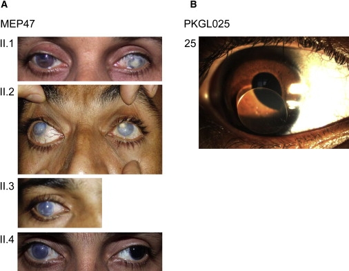
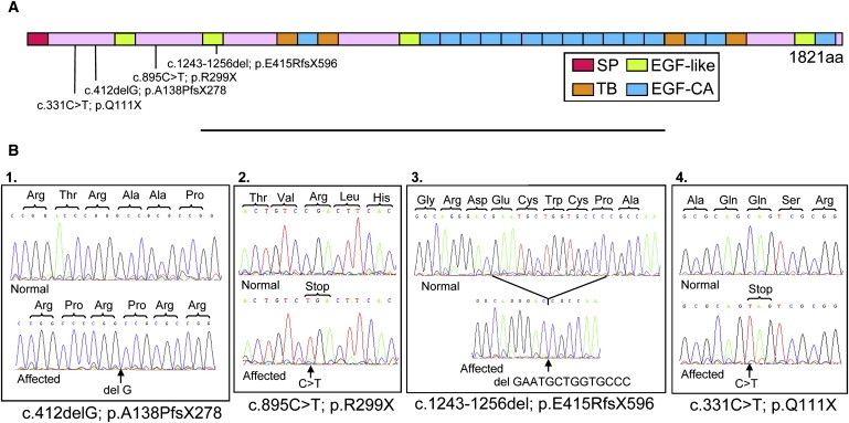
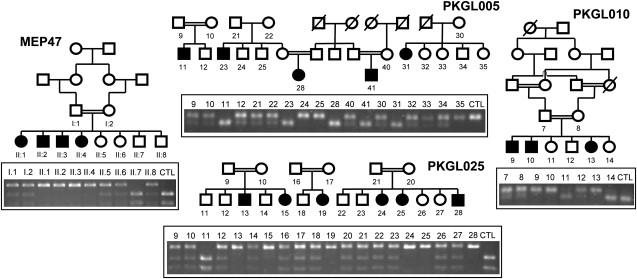
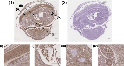
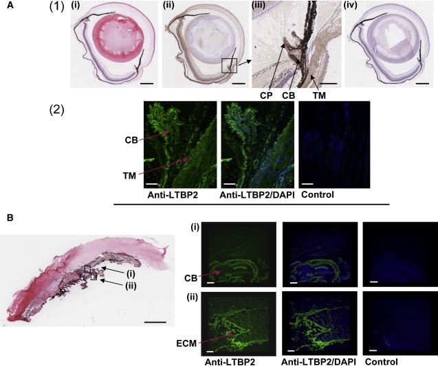
Primary congenital glaucoma (PCG) is an autosomal-recessive condition characterized by high intraocular pressure (IOP), usually within the first year of life, which potentially could lead to optic nerve damage, globe enlargement, and permanent loss of vision. To date, PCG has been linked to three loci: 2p21 (GLC3A), for which the responsible gene is CYP1B1, and 1p36 (GLC3B) and 14q24 (GLC3C), for which the genes remain to be identified. Here we report that null mutations in LTBP2 cause PCG in four consanguineous families from Pakistan and in patients of Gypsy ethnicity. LTBP2 maps to chromosome 14q24.3 but is around 1.3 Mb proximal to the documented GLC3C locus. Therefore, it remains to be determined whether LTBP2 is the GLC3C gene or whether a second adjacent gene is also implicated in PCG. LTBP2 is the largest member of the latent transforming growth factor (TGF)-beta binding protein family, which are extracellular matrix proteins with multidomain structure. It has homology to fibrillins and may have roles in cell adhesion and as a structural component of microfibrils. We confirmed localization of LTBP2 in the anterior segment of the eye, at the ciliary body, and particularly the ciliary process. These findings reveal that LTBP2 is essential for normal development of the anterior chamber of the eye, where it may have a structural role in maintaining ciliary muscle tone.


Figures

Figure 1
Figure 1
Clinical Features of PCG Patients (A) For the MEP47 family, photos of the anterior segment of the affected family members are shown. At the time of examination, patients II.1, II.2, II.3, and II.4 were aged 42, 34, 26, and 24 years old, respectively. Case II.1 has Haab's striae in the right eye and a central corneal scar secondary to infection in the left eye. Cases II.2, II.3 (right eye), and II.4 (right eye) have corneal enlargement and clouding. The left eye of case II.4 has an enlarged but clear cornea after surgery. (B) Slit lamp examination of an affected patient 25 aged 5 years old from family PKGL025 shows dislocation of the lens (ectopia lentis).
Figure 2
Figure 2
LTBP2 Genetic Analysis in PCG Affecteds (A) Diagrammatic representation of the LTBP2 protein illustrating the position of the four mutations relative to the full-length 1821 amino acid protein. The domains in the protein are also shown; SP depicts the signal peptide, TB the transforming growth factor beta binding protein-like motif, EGF-like and EGF-CA both represent epidermal growth factor-like domains but the latter also has a calcium binding motif. (B) Sequence chromatograms of a normal individual and one PCG-affected patient from each of the families. The sequences highlight the mutation in (1) MEP47 (exon 1), (2) PKGL005 (exon 4), (3) PKGL025 (exon 6), and (4) PKGL010 (exon 1).
Figure 3
Figure 3
Segregation of the LTBP2 Mutation in Each of the Families The pedigree structures and individual IDs are shown for MEP47 and PKGL010; however, only the families from which DNA is available are shown for PKGL005 and PKGL025 because the full pedigrees have been published before. The agarose gel photographs depicted show segregation of all four mutations with the disease phenotype in each of the respective families as would be expected for a recessive mode of inheritance. Only the PCG-affected members have both LTBP2 alleles mutated as illustrated by the unique banding pattern on the agarose gel. The banding pattern for CTL represents a wild-type control individual.
Figure 4
Figure 4
Immunohistological Localization of LTBP2 in Mouse Embryonic Tissue Section of whole mouse embryo at E15 stained with (1) rabbit polyclonal LTBP2 antibody or (2) the Rabbit Envision Detection system as negative control. LTBP2 immunoreactivity depicted as brown staining was present in the connective tissues throughout the body with an increased localization in the spinal cord (i), the cardiac muscle (ii), the skeletal muscle (iii), and the renal and seminiferous tubules (iv). Scale bars represent 500 μm.
Figure 5
Figure 5
Localization of LTBP2 in the Adult Eye (A) (1) Formalin-fixed sections of adult mouse eyes stained with hematoxylin and eosin (i), anti-LTBP2 (ii), or the Rabbit Envision Detection system (iv). In (ii), LTBP2 immunoreactivity was observed as brown staining in several structures of the eye including the sclera, retina, cornea, trabecular meshwork, and the ciliary body. The black intense pigment that extends from the retinal pigment epithelium to the iris is melanin (in (i), (ii), and (iv)). Scale bars represent 500 μm. The area surrounding the ciliary body in (ii) has been magnified and is shown in (iii). Note LTBP2 immunoreactivity particularly at the trabecular meshwork (TM), the ciliary body (CB), and the ciliary process (CP). Scale bar represents 100 μm. (2) Cryopreserved sections of adult mouse eyes stained either with anti-LTBP2 or a rabbit IgG isotype negative control. LTBP2 immunoreactivity depicted as green fluorescence was present in the trabecular meshwork (TM), the ciliary body (CB), and particularly the ciliary process. Scale bars represent 50 μm. (B) Cow eye histological section showing the cornea, iris, and ciliary body stained with hematoxylin and eosin. Scale bar represents 1500 μm. The lens dislocated during the sectioning and the adjacent alignment of the iris with the cornea in the panel is an artifact of tissue preparation. Cryosections stained with anti-LTBP2 or rabbit IgG negative control are shown. Note that the green fluorescence corresponding to LTBP2 immunoreactivity is abundant in the ciliary body (CB) (i) and particularly intense in the extracellular matrix structures (ECM) at the ciliary process (ii). Scale bars represent 150 μm.

References

    1. Kupfer C., Kaiser-Kupfer M.I. Observations on the development of anterior chamber angle with reference to the pathogenesis of congenital glaucomas. Am. J. Ophthalmol. 1979;88:424–426. - PubMed
    1. Anderson D.R. The development of the trabecular meshwork and its abnormality in primary infantile glaucoma. Trans. Am. Ophthalmol. Soc. 1981;79:458–485. - PMC - PubMed
    1. Ho C.L., Walton D.S. Primary congenital glaucoma: 2004 update. J. Pediatr. Ophthalmol. Strabismus. 2004;41:271–288. - PubMed
    1. Francois J. Congenital glaucoma and its inheritance. Ophthalmologica. 1980;181:61–73. - PubMed
    1. Dandona L., Williams J.D., Williams B.C., Rao G.N. Population-based assessment of childhood blindness in Southern India. Arch. Ophthalmol. 1998;116:545–546. - PubMed

Publication types

Substances