

Node of Ranvier formation on motoneurons in vitro
- PMID: 19361859
- PMCID: PMC3793346
- DOI: 10.1016/j.biomaterials.2009.03.023
Node of Ranvier formation on motoneurons in vitro
Abstract
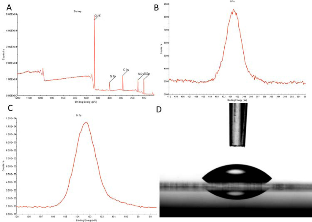
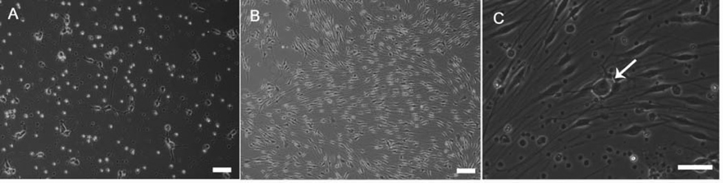
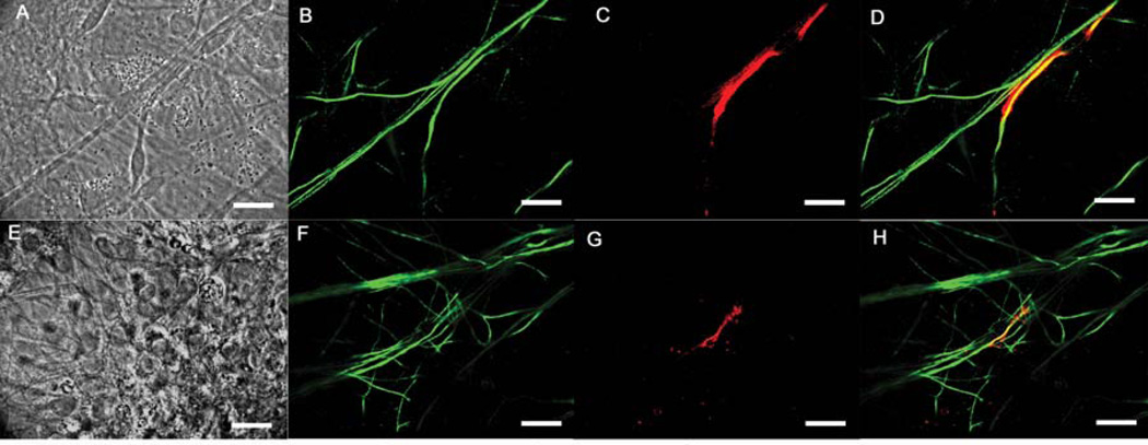
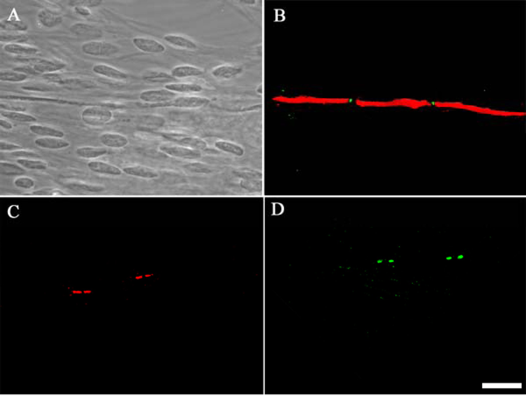
One of the most significant interactions between Schwann cells and neurons is myelin sheath formation. Myelination is a vertebrate adaptation that enables rapid conduction of action potentials without a commensurate increase in axon diameter. In vitro neuronal systems provide a unique modality to study both factors influencing myelination and diseases associated with myelination. Currently, no in vitro system for motoneuron myelination by Schwann cells has been demonstrated. This work details the myelination of motoneuron axons by Schwann cells, with complete Node of Ranvier formation, in a defined in vitro culture system. This defined system utilizes a novel serum-free medium in combination with the non-biological substrate, N-1[3 (trimethoxysilyl) propyl] diethylenetriamine (DETA). The myelinated segments and nodal proteins were visualized and quantified using confocal microscopy. This defined system provides a highly controlled, reproducible model for studying Schwann cell interactions with motoneurons as well as the myelination process and its effect on neuronal plasticity. Furthermore, an in vitro system that would allow studies of motoneuron myelination would be beneficial for understanding peripheral demyelinating neuropathies such as diabetes induced peripheral neuropathy and could lead to a better understanding of CNS demyelinating diseases like multiple sclerosis, as well as neuromuscular junction maturation and maintenance.
Figures
Similar articles
-
Myelination and node of Ranvier formation on sensory neurons in a defined in vitro system.In Vitro Cell Dev Biol Anim. 2013 Sep;49(8):608-618. doi: 10.1007/s11626-013-9647-8. Epub 2013 Aug 16. In Vitro Cell Dev Biol Anim. 2013. PMID: 23949775 Free PMC article.
-
Establishment of a Serum-Free Human iPSC-Derived Model of Peripheral Myelination.ACS Biomater Sci Eng. 2024 Nov 11;10(11):7132-7143. doi: 10.1021/acsbiomaterials.4c01431. Epub 2024 Oct 22. ACS Biomater Sci Eng. 2024. PMID: 39437333 Free PMC article.
-
Nodes of Ranvier form in association with ezrin-radixin-moesin (ERM)-positive Schwann cell processes.Proc Natl Acad Sci U S A. 2001 Jan 30;98(3):1235-40. doi: 10.1073/pnas.98.3.1235. Proc Natl Acad Sci U S A. 2001. PMID: 11158623 Free PMC article.
-
Molecular organization and function of vertebrate septate-like junctions.Biochim Biophys Acta Biomembr. 2020 May 1;1862(5):183211. doi: 10.1016/j.bbamem.2020.183211. Epub 2020 Feb 4. Biochim Biophys Acta Biomembr. 2020. PMID: 32032590 Review.
-
The local differentiation of myelinated axons at nodes of Ranvier.Nat Rev Neurosci. 2003 Dec;4(12):968-80. doi: 10.1038/nrn1253. Nat Rev Neurosci. 2003. PMID: 14682359 Review.
Cited by
-
A phenotypic culture system for the molecular analysis of CNS myelination in the spinal cord.Biomaterials. 2014 Oct;35(31):8840-8845. doi: 10.1016/j.biomaterials.2014.07.007. Epub 2014 Jul 23. Biomaterials. 2014. PMID: 25064806 Free PMC article.
-
Myelination and node of Ranvier formation on sensory neurons in a defined in vitro system.In Vitro Cell Dev Biol Anim. 2013 Sep;49(8):608-618. doi: 10.1007/s11626-013-9647-8. Epub 2013 Aug 16. In Vitro Cell Dev Biol Anim. 2013. PMID: 23949775 Free PMC article.
-
A phenotypic in vitro model for the main determinants of human whole heart function.Biomaterials. 2015 Aug;60:20-30. doi: 10.1016/j.biomaterials.2015.04.035. Epub 2015 May 14. Biomaterials. 2015. PMID: 25978005 Free PMC article.
-
Neuromuscular junction formation between human stem cell-derived motoneurons and human skeletal muscle in a defined system.Biomaterials. 2011 Dec;32(36):9602-11. doi: 10.1016/j.biomaterials.2011.09.014. Epub 2011 Sep 23. Biomaterials. 2011. PMID: 21944471 Free PMC article.
-
A Novel Bioengineered Functional Motor Unit Platform to Study Neuromuscular Interaction.J Clin Med. 2020 Oct 10;9(10):3238. doi: 10.3390/jcm9103238. J Clin Med. 2020. PMID: 33050427 Free PMC article.
References
-
- Jessen KR, Mirsky R. The origin and development of glial cells in peripheral nerves. Nat Rev Neurosci. 2005;6(9):671–682. - PubMed
-
- Sherman DL, Tait S, Melrose S, Johnson R, Zonta B, Court FA, et al. Neurofascins Are Required to Establish Axonal Domains for Saltatory Conduction. Neuron. 2005;48(5):737. - PubMed
-
- Sherman DL, Brophy PJ. Mechanisms of axon ensheathment and myelin growth. Nat Rev Neurosci. 2005;6(9):683–690. - PubMed
-
- Wood P, Moya F, Eldridge CF, Owens G, Ranscht B, Schachner M, et al. Studies of the initiation of myelination by Schwann cells. Ann N Y Acad Sci. 1990;605:1–14. - PubMed
-
- Bahr M, Hopkins JM, Bunge RP. In vitro myelination of regenerating adult rat retinal ganglion cell axons by Schwann cells. Glia. 1991;4(5):529–533. - PubMed
Publication types
MeSH terms
Grants and funding
LinkOut - more resources
Full Text Sources
Other Literature Sources
