. 2009 Jul;30(20):3405-14.
doi: 10.1016/j.biomaterials.2009.02.046. Epub 2009 Apr 9.

Engineering retinal progenitor cell and scrollable poly(glycerol-sebacate) composites for expansion and subretinal transplantation

Affiliations

Engineering retinal progenitor cell and scrollable poly(glycerol-sebacate) composites for expansion and subretinal transplantation

Stephen Redenti et al. Biomaterials. 2009 Jul.

Abstract

Retinal degenerations cause permanent visual loss and affect millions world-wide. Presently, a novel treatment highlights the potential of using biodegradable polymer scaffolds to induce differentiation and deliver retinal progenitor cells for cell replacement therapy. In this study, we engineered and analyzed a micro-fabricated polymer, poly(glycerol sebacate) (PGS) scaffold, whose useful properties include biocompatibility, elasticity, porosity, and a microtopology conducive to mouse retinal progenitor cell (mRPC) differentiation. In vitro proliferation assays revealed that PGS held up to 86,610 (+/-9993) mRPCs per square millimeter, which were retained through simulated transplantations. mRPCs adherent to PGS differentiated toward mature phenotypes as evidenced by changes in mRNA, protein levels, and enhanced sensitivity to glutamate. Transplanted composites demonstrated long-term mRPC survival and migrated cells exhibited mature marker expression in host retina. These results suggest that combining mRPCs with PGS scaffolds for subretinal transplantation is a practical strategy for advancing retinal tissue engineering as a restorative therapy.


Figures

Fig. 1
Fig. 1
Retinal transplantation ex vivo method. a) GFP + mRPCs are seeded and allowed to proliferate for one week on PGS in a culture insert containing 0.4 µm pores which provides a restricted surface area and allows for the exchange of nutrients from culture media contained in the surrounding culture well. b) At day 7, freshly isolated retina are explanted onto a culture well insert with ganglion cell layer (GLC) at bottom, inner nuclear layer (INL) center and outer nuclear layer (ONL) at top. PGS–mRPC composites are then transferred to the ONL surface and allowed to culture and migrate into retinal lamina for 7 days.
Fig. 2
Fig. 2
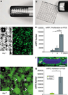
Proliferation and adherence of mRPCs cultured on PGS. a) Elastic and porous PGS scrolled within a 1 mm inner diameter glass needle and d) injected and unscrolled (scale, 1 mm). b) Day 1 post-GFP+ mRPC seeded PGS shown left under white light and right under fluorescent illumination. c) GFP+ mRPCs fully cover PGS with proliferative neurospheres (arrows) by day 7 (scale b,c, 100 µm). e) mRPC numbers on 1 ×1 mm PGS sections increased from 9077 (±4748), 20,003 (±10,223) and 86,610 (±9933) at days 1, 3 and 7 respectively. (days 1–7, *p = .002, days 3–7, *p = .004) f) 40× section of GFP mRPCs adherent to PGS (pseudo color blue) on upper and lower surfaces and through pores (scale, 50 µm). g) Following identical treatment and mRPC seeding density (2.5 × 10⁵), 2× 2 mm PGS retained a significantly higher number of cells than glass after 7 days in culture transplantation simulation 229,992 (±68,022) and 15,596 (±12,418), respectively (*p= 0.03). Error bars, s.e.m.
Fig. 3
Fig. 3
SEM of PGS topology and mRPC adhesion. a) Top view of PGS 45 µm thick PGS scaffold with 50 µm diameter pores spaced 175 µm apart. b) Top view of PGS seeded with mRPCs after 7 days of proliferation surrounding pore (circle). c) Magnification of b showing individual mRPCs with flattened radial and bipolar morphology. d) Side view of PGS scaffold showing the cone-like pore formation on the upper surface and 45 µm thickness. e) Adhesion of neurospheres to the PGS surface, along the sidewalls, and within individual pores (circle). f) Magnification of e showing spheroid mRPC infiltration into an individual pore.
Fig. 4
Fig. 4
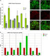
Immunocytochemical and qPCR analysis of PGS influenced expression levels. To evaluate the influence of the PGS microenvironment on cell differentiation, intensity of immunocytochemical labeling was compared between mRPCs cultured on either 1 × 1 mm PGS or glass for 7 days. a) Marker expression indicating undifferentiated mRPCs including Pax6, Hes1, nestin, and Sox2 was decreased in cells cultured on PGS. Elevated GFAP expression may indicate a higher percentage of mRPCs of glial fate on glass than on PGS. b,e) GFP+ mRPCs expressing c,f) nestin were analyzed in overlaid images d,g) for percent expression between glass (b–d) and PGS (e–g) samples. Cells cultured on PGS expressed lower levels of nestin. h) Results from quantitative PCR analysis showed that message expression correlated well with protein data, the exception being an increase in the level of nestin message in response to growth on PGS (b–g) (green = GFP, red = rhodamine labeled protein).
Fig. 5
Fig. 5
mRPCs respond to glutamate, NMDA + glycine with Ca2+ influxes. a,b) Averaged responses of Fura-2 loaded PGS–mRPCs (n = 25) demonstrate a 40% greater peak intra-cellular Ca2+ influx above glass–mRPCs (n = 25) in response to 1 mm glutamate. c) Pseudo color images illustrating 340:380 ratio levels of Fura-2 loaded mRPCs before (left) and at the peak of glutamate stimulation (right) (scale, 50 µm). d) Responses of mRPCs stimulated with 1 mm glutamate,1 mm NMDA with 1 mm glycine. Averaged glutamate responses by mRPCs on PGS (max avg: 641.16 nm, 39.78 s.e.m., n = 25) were significantly higher than those on glass (max avg: 458.92 nm, 37.42 s.e.m., n = 25, **p = 0.001) Responses to NMDA with glycine were not significantly different between groups.
Fig. 6
Fig. 6
PGS scaffold delivery of GFP+ mRPCs to C57bl/6 and rho−/− mouse retinal explants. Scaffolds were seeded with ~2.5 × 10⁵ at day P0 with GFP+ mRPCs (green) and allowed to proliferate in vitro for 7 days. a–e) C57bl/6 retina. a) GFP+ mRPCs co-labeled (yellow) for nf-200 (arrows), delivered on PGS aligned with the ONL from which cells extended processes and migrated into each retinal layer. b) PGS delivered mRPCs label for nestin in the ONL, c) NeuN (neuronal nuclei) in the ONL, d) PKC in the INL region, and e) GFAP in the GCL region. Antibody staining is shown in red. f–j) rho−/− retina. The ONL has degenerated in these rho−/− retina and PGS with attached layer of mRPCs aligned with the INL. mRPCs migrated into each of the remaining inner nuclear, inner plexiform and ganglion cell layer. f) GFP+ mRPCs that reached the GCL layer expressed GFAP (arrows). g) mRPCs still attached to the PGS scaffold labeled for NeuN, h) cells that reached the INL expressed crx, i) nestin in the IPL region, and j) GFAP in the GCL region (green = GFP, red = rhodamine labeled marker, blue = Topro-3 nuclei label) scale, 25 µm.
Fig. 7
Fig. 7
GFP+ mRPC migration and differentiation in C57bl/6 retina 30 days following sub-retinal transplantation. a) PGS delivered GFP+ mRPCs (green) migrate into the inner plexiform layer and co-label for GFAP (red). Inset a) shows a PGS–RPC composite scrolled and loaded (left) into a 1 mm I.D glass tube and ejected (right) unrolled to present its cell cargo (scale, 1 mm). Small mRPCs migrated into the ONL label for b) crx, c) in NeuN, and d) rhodopsin. In the inner nuclear layer migrated cells label for PKC (scale, 50 µm). f) At day 7, the number of mRPCs migrated into 18 µm thick C57bl/6 and rho−/− retinal explant sections were 175 ± 57 and 94 ± 33, respectively. g) The number of mRPCs in each 18 µm section of C57bl/6 and rho−/− retina at 30 days post-transplantation was 28 ± 8 and 15 ± 4, respectively (green = GFP, red = rhodamine labeled protein, blue = Topro-3 nuclei label).

References

    1. Margalit E, Sadda SR. Retinal and optic nerve diseases. Artif Organs. 2003;27(11):963–974. - PubMed
    1. Klassen HJ, Ng TF, Kurimoto Y, Kirov I, Shatos M, Coffey P, et al. Multipotent retinal progenitors express developmental markers, differentiate into neurons, and preserve light-mediated behavior. Invest Ophthalmol Vis Sci. 2004;45(11):4167–4173. - PubMed
    1. MacLaren RE, Pearson RA, MacNeil A, Douglas RH, Salt TE, Akimoto M, et al. Retinal repair by transplantation of photoreceptor precursors. Nature. 2006;444(7116):203–207. - PubMed
    1. Tomita M, Lavik E, Klassen H, Zahir T, Langer R, Young MJ. Biodegradable polymer composite grafts promote the survival and differentiation of retinal progenitor cells. Stem Cells. 2005;23(10):1579–1588. - PubMed
    1. Tao S, Young C, Redenti S, Zhang Y, Klassen H, Desai T, et al. Survival, migration and differentiation of retinal progenitor cells transplanted on micro-machined poly(methyl methacrylate) scaffolds to the subretinal space. Lab Chip. 2007;7(6):695–701. - PubMed

Publication types

MeSH terms