

Retinotopically specific reorganization of visual cortex for tactile pattern recognition
- PMID: 19361999
- PMCID: PMC2709730
- DOI: 10.1016/j.cub.2009.02.063
Retinotopically specific reorganization of visual cortex for tactile pattern recognition
Abstract
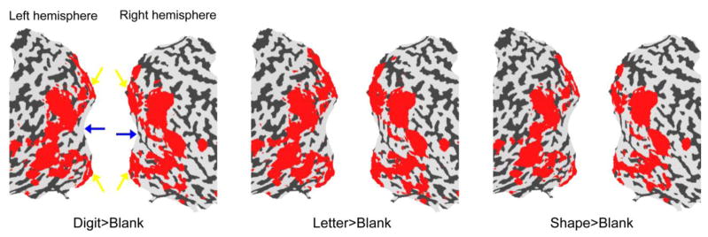
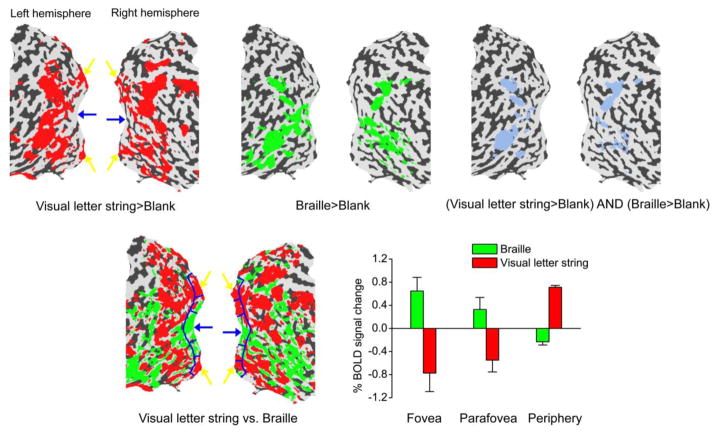
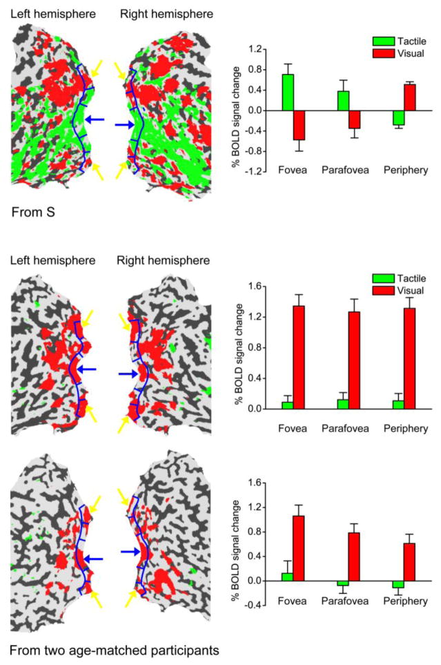
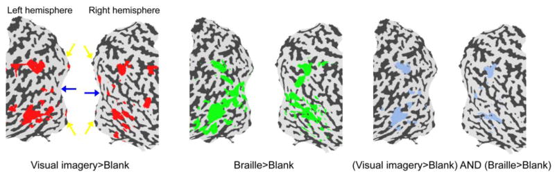
Although previous studies have shown that Braille reading and other tactile discrimination tasks activate the visual cortex of blind and sighted people, it is not known whether this kind of crossmodal reorganization is influenced by retinotopic organization. We have addressed this question by studying "S," a visually impaired adult with the rare ability to read print visually and Braille by touch. S had normal visual development until 6 years of age, and thereafter severe acuity reduction due to corneal opacification, but no evidence of visual-field loss. Functional magnetic resonance imaging revealed that, in S's early visual areas, tactile information processing activated what would be the foveal representation for normally sighted individuals, and visual information processing activated what would be the peripheral representation. Control experiments showed that this activation pattern was not due to visual imagery. S's high-level visual areas, which correspond to shape- and object-selective areas in normally sighted individuals, were activated by both visual and tactile stimuli. The retinotopically specific reorganization in early visual areas suggests an efficient redistribution of neural resources in the visual cortex.
Figures
Similar articles
-
In a case of longstanding low vision regions of visual cortex that respond to tactile stimulation of the finger with Braille characters are not causally involved in the discrimination of those same Braille characters.Cortex. 2022 Oct;155:277-286. doi: 10.1016/j.cortex.2022.07.012. Epub 2022 Aug 11. Cortex. 2022. PMID: 36054997
-
Recruitment of Foveal Retinotopic Cortex During Haptic Exploration of Shapes and Actions in the Dark.J Neurosci. 2017 Nov 29;37(48):11572-11591. doi: 10.1523/JNEUROSCI.2428-16.2017. Epub 2017 Oct 24. J Neurosci. 2017. PMID: 29066555 Free PMC article.
-
Orthographic Priming in Braille Reading as Evidence for Task-specific Reorganization in the Ventral Visual Cortex of the Congenitally Blind.J Cogn Neurosci. 2019 Jul;31(7):1065-1078. doi: 10.1162/jocn_a_01407. Epub 2019 Apr 2. J Cogn Neurosci. 2019. PMID: 30938589
-
Feeling with the mind's eye: contribution of visual cortex to tactile perception.Behav Brain Res. 2002 Sep 20;135(1-2):127-32. doi: 10.1016/s0166-4328(02)00141-9. Behav Brain Res. 2002. PMID: 12356442 Review.
-
Cross-modal plasticity of tactile perception in blindness.Restor Neurol Neurosci. 2010;28(2):271-81. doi: 10.3233/RNN-2010-0534. Restor Neurol Neurosci. 2010. PMID: 20404414 Free PMC article. Review.
Cited by
-
Perceptual learning modifies the functional specializations of visual cortical areas.Proc Natl Acad Sci U S A. 2016 May 17;113(20):5724-9. doi: 10.1073/pnas.1524160113. Epub 2016 Apr 5. Proc Natl Acad Sci U S A. 2016. PMID: 27051066 Free PMC article.
-
Low Vision and Plasticity: Implications for Rehabilitation.Annu Rev Vis Sci. 2016 Oct 14;2:321-343. doi: 10.1146/annurev-vision-111815-114344. Epub 2016 Jul 25. Annu Rev Vis Sci. 2016. PMID: 28532346 Free PMC article. Review.
-
Dance Is More Than Meets the Eye-How Can Dance Performance Be Made Accessible for a Non-sighted Audience?Front Psychol. 2021 Apr 16;12:643848. doi: 10.3389/fpsyg.2021.643848. eCollection 2021. Front Psychol. 2021. PMID: 33935898 Free PMC article.
-
Correlation of vision loss with tactile-evoked V1 responses in retinitis pigmentosa.Vision Res. 2015 Jun;111(Pt B):197-207. doi: 10.1016/j.visres.2014.10.015. Epub 2014 Nov 3. Vision Res. 2015. PMID: 25449160 Free PMC article.
-
Interplay between Heightened Temporal Variability of Spontaneous Brain Activity and Task-Evoked Hyperactivation in the Blind.Front Hum Neurosci. 2016 Dec 20;10:632. doi: 10.3389/fnhum.2016.00632. eCollection 2016. Front Hum Neurosci. 2016. PMID: 28066206 Free PMC article.
References
-
- Sadato N, Pascual-Leone A, Grafman J, Ibanez V, Deiber MP, Dold G, Hallett M. Activation of the primary visual cortex by Braille reading in blind subjects. Nature. 1996;380:526–528. - PubMed
-
- Buchel C, Price C, Frackowiak RS, Friston K. Different activation patterns in the visual cortex of late and congenitally blind subjects. Brain. 1998;121:409–419. - PubMed
-
- Cohen LG, Weeks RA, Sadato N, Celnik P, Ishii K, Hallett M. Period of susceptibility for cross-modal plasticity in the blind. Ann Neurol. 1998;45:451–460. - PubMed
-
- Sadato N, Okada T, Honda M, Yonekura Y. Critical period for cross-modal plasticity in blind humans: a functional MRI study. NeuroImage. 2002;16:389–400. - PubMed
Publication types
MeSH terms
Grants and funding
LinkOut - more resources
Full Text Sources
