. 2009 Apr 17;30(4):566-75.
doi: 10.1016/j.immuni.2009.02.006. Epub 2009 Apr 9.

The intracellular sensor NLRP3 mediates key innate and healing responses to influenza A virus via the regulation of caspase-1

Affiliations

The intracellular sensor NLRP3 mediates key innate and healing responses to influenza A virus via the regulation of caspase-1

Paul G Thomas et al. Immunity. .

Abstract

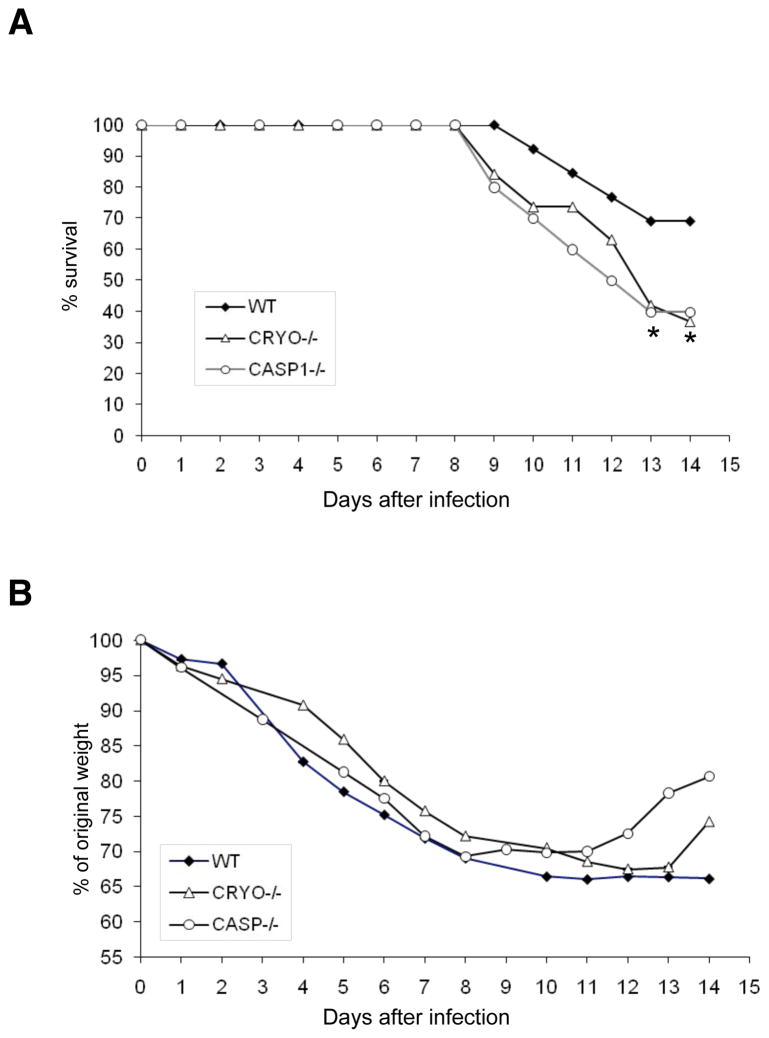
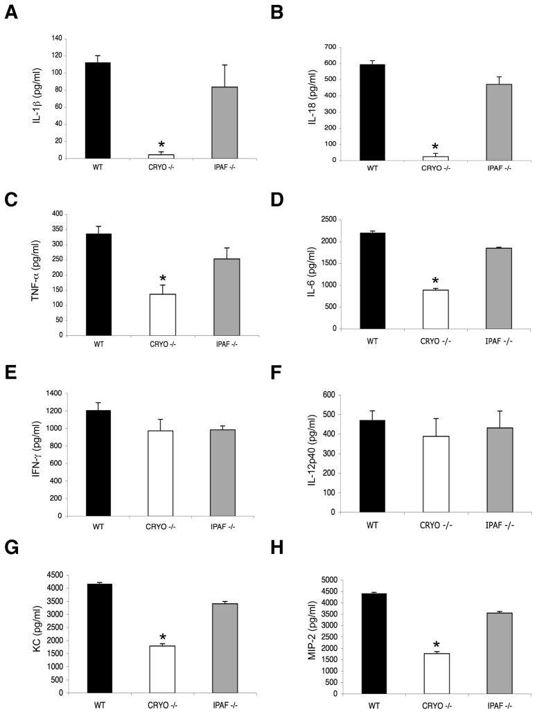
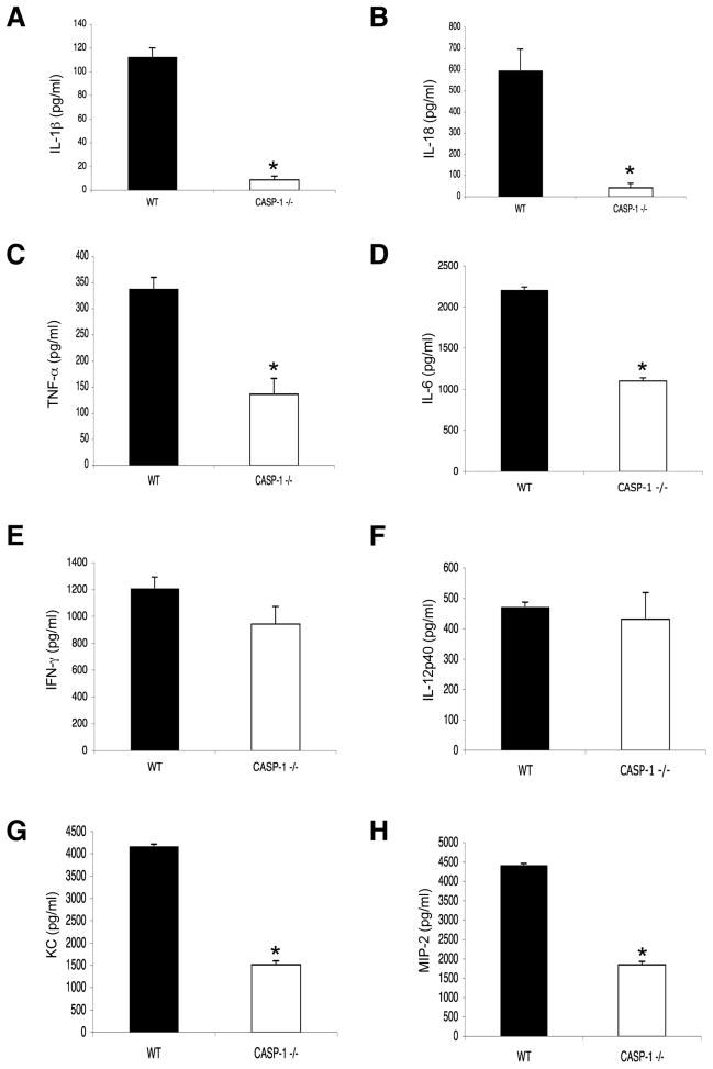
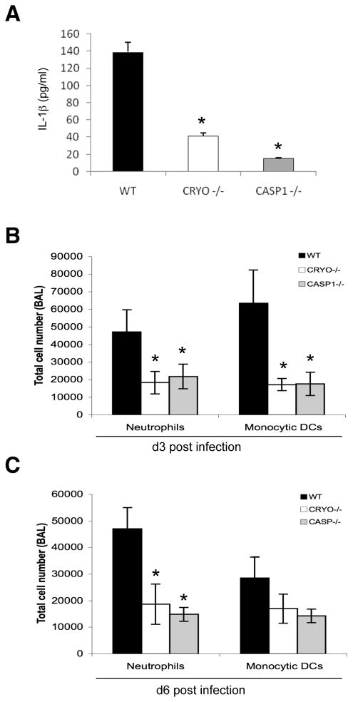
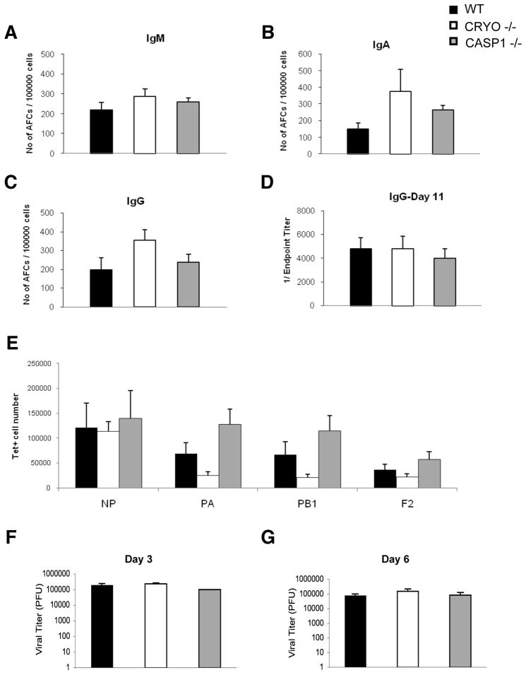
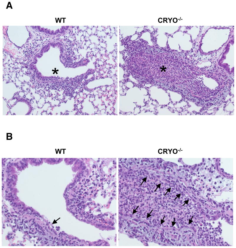
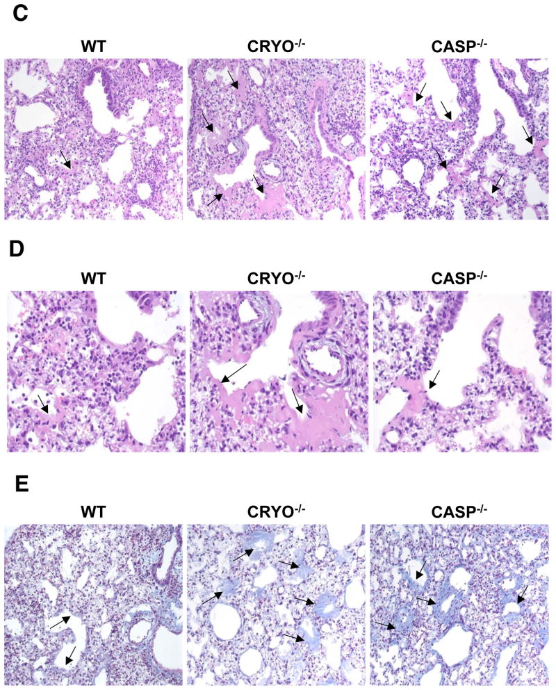
Virus-induced interlukin-1beta (IL-1beta) and IL-18 production in macrophages are mediated via caspase-1 pathway. Multiple microbial components, including viral RNA, are thought to trigger assembly of the cryopyrin inflammasome resulting in caspase-1 activation. Here, we demonstrated that Nlrp3(-/-) and Casp1(-/-) mice were more susceptible than wild-type mice after infection with a pathogenic influenza A virus. This enhanced morbidity correlated with decreased neutrophil and monocyte recruitment and reduced cytokine and chemokine production. Despite the effect on innate immunity, cryopyrin-deficiency was not associated with any obvious defect in virus control or on the later emergence of the adaptive response. Early epithelial necrosis was, however, more severe in the infected mutants, with extensive collagen deposition leading to later respiratory compromise. These findings reveal a function of the cryopyrin inflammasome in healing responses. Thus, cryopyrin and caspase-1 are central to both innate immunity and to moderating lung pathology in influenza pneumonia.


Figures

Figure 1
Figure 1. Cryopyrin and caspase-1 provide protection against influenza A virus-induced lethality
Groups of 10 wildtype, cryopyrin−/− (CRYO−/−) and casapse-1 (CASP1−/−) mice were infected i.n. with 8×10³ EID50 (= 1LD50) of PR8 and survival was monitored daily for 14d. Differences in group survival were analyzed with Cox proportional hazards test and p<0.05 was considered statistically significant.
Figure 2
Figure 2. Influenza virus-induced cytokine and chemokine production depends on cryopyrin, but not ipaf
Groups (n=4) of B6 (WT), cryopyrin−/− (CRYO−/−) and ipaf−/− (IPAF−/−) mice were infected i.n with 4×10³ EID50 of PR8 influenza virus and sampled 3d later to determine levels of IL-1β (A), IL-18 (B), TNF-α (C), IL-6 (D), IFN-γ (E), IL-12p40 (F), KC (G), and MIP-2 (H), in BAL fluid. Results are mean ± S.D. and are representative of three independent experiments. Data were analyzed with Student’s t-test (*, p<0.05).
Figure 3
Figure 3. Caspase-1 is required for influenza virus-induced secretion of cryopyrin-dependent cytokines and chemokines
Groups of 4 B6 (WT) and caspase-1−/− (CASP1−/−) mice were infected i.n with 4×10³ EID50 of PR8 influenza virus and levels of IL-1β (A), IL-18 (B), TNF-α (C), IL-6 (D), IFN-γ (E), IL-12p40 (F), KC (G), and MIP-2 (H) were measured in BAL fluid 3d later. Results are mean ± S.D.. Data were analyzed with Student’s t-test (*, p<0.05) and are representative of three independent experiments.
Figure 4
Figure 4. Innate cellular responses to influenza are mediated by cryopyrin and caspase-1
(A) Dendritic cells from WT, CRYO−/− and CASP1−/− mice were transfected with 2 μg purified influenza A viral RNA for 24 h and cell-free supernatants were analyzed by ELISA for production of IL-1β. Data are representative of two independent experiments. Groups of 4 C57BL/6 WT, CRYO−/− and CASP1−/− mice were infected i.n. with 2×10³ EID50 of PR8 influenza virus and cells recovered by BAL on d3 (B) and d6 (C) were characterized by flow cytometry (see Experimental Procedures). Results are mean ± S.D. Data were analyzed with ANOVA (*, p<0.05) and are representative of three independent experiments.
Figure 5
Figure 5. Cryopyrin and caspase-1 are not required for early virus clearance or for adaptive immunity
Groups of 4 WT, CRYO −/− and CASP1−/− mice were infected i.n. with 4×10³ EID50 of PR8 influenza. Seven days following infection, the frequency of antibody secreting cells in the draining mediastinal lymph node was determined by ELISPOT for the indicated isotype (A–C). Antigen-specific serum IgG was measured by ELISA on d11 following infection (D). Also on d11, antigen-specific CD8+ T cell numbers were enumerated from the BAL by tetramer staining (E). Virus titers in lungs were determined on d3 (F) and d6 (G) by standard MDCK plaque assay. Data were analyzed with ANOVA (*, p<0.05) and are representative of three independent experiments.
Figure 6
Figure 6. Virus-induced pulmonary necrosis is enhanced in the absence of cryopyrin
Groups of 4 WT and cryopyrin−/− mice were infected i.n. with 8×10³ EID50 of PR8 influenza virus. Lung samples were collected on d3 and processed for routine H&E staining of 4μ paraffin sections. (A), top panels, ×200, WT airway epithelium is intact with only small foci of necrosis (arrow) and peribronchiolar inflammation; * shows unobstructed airway lumen. Cryopyrin−/−, the small bronchi and bronchioles show diffuse necrosis with loss of the airway epithelium and obstruction of the lumen by debris (*). (B), bottom panels ×400, Intact wildtype epithelium with limited foci of necrosis (arrow), and diffuse necrosis of CRYO−/− bronchiolar epithelium (arrows). Histologic evaluation was performed by an experienced veterinary pathologist (KLB). Groups of 4 wildtype and cryopyrin−/− mice were infected i.n. with 8×10³ EID50 of PR8 influenza virus. Lung samples were collected on d11 and 4μm paraffin sections processed for routine H&E (C,D) or Masson’s Trichrome staining (E). (C) ×200, the WT mice show only focal and minimal collagen deposition in the alveoli and occasionally in the terminal airways (arrows). This collagen deposition is exuberant in the mutant groups and often occludes alveoli and terminal airways (arrows). (D) ×400, arrows indicate areas of collagen deposition. (E) ×200, bottom row Masson’s Trichrome stain confirms collagen deposition. In the WT groups the insult was sufficiently severe to cause basement membrane damage, evidenced by a thin layer of collagen lining the alveolar spaces. In the mutant lungs, deposition of collagen is abundant and forms concentric layers that occlude alveoli and terminal airways.

Similar articles

Cited by

References

    1. Allan W, Tabi Z, Cleary A, Doherty PC. Cellular events in the lymph node and lung of mice with influenza. Consequences of depleting CD4+ T cells. J Immunol. 1990;144:3980–3986. - PubMed
    1. Amer A, Franchi L, Kanneganti TD, Body-Malapel M, Ozoren N, Brady G, Meshinchi S, Jagirdar R, Gewirtz A, Akira S, et al. Regulation of Legionella phagosome maturation and infection through flagellin and host IPAF. J Biol Chem 2006 - PubMed
    1. Burns K, Martinon F, Tschopp J. New insights into the mechanism of IL-1beta maturation. Curr Opin Immunol. 2003;15:26–30. - PubMed
    1. de Jong MD, Simmons CP, Thanh TT, Hien VM, Smith GJ, Chau TN, Hoang DM, Chau NV, Khanh TH, Dong VC, et al. Fatal outcome of human influenza A (H5N1) is associated with high viral load and hypercytokinemia. Nat Med. 2006;12:1203–1207. - PMC - PubMed
    1. Denton AE, Doherty PC, Turner SJ, La Gruta NL. IL-18, but not IL-12, is required for optimal cytokine production by influenza virus-specific CD8+ T cells. Eur J Immunol. 2007;37:368–375. - PubMed

Publication types