

Localization of myocyte enhancer factor 2 in the rodent forebrain: regionally-specific cytoplasmic expression of MEF2A
- PMID: 19362076
- PMCID: PMC2723059
- DOI: 10.1016/j.brainres.2009.03.067
Localization of myocyte enhancer factor 2 in the rodent forebrain: regionally-specific cytoplasmic expression of MEF2A
Abstract
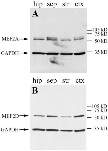
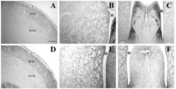
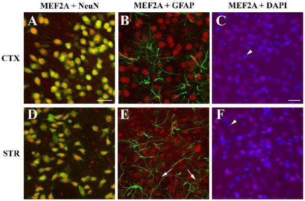
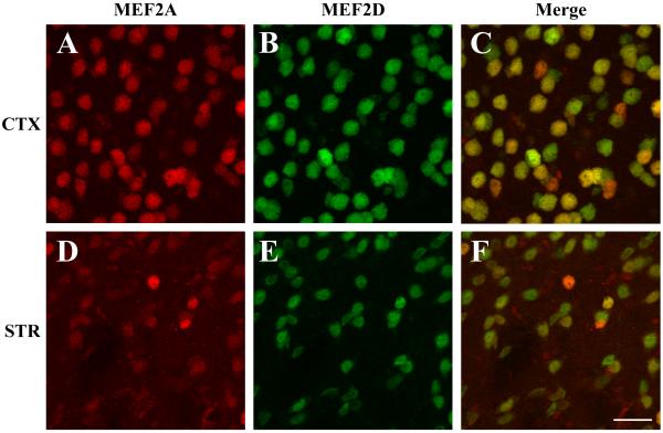
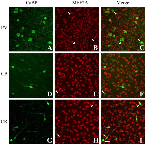
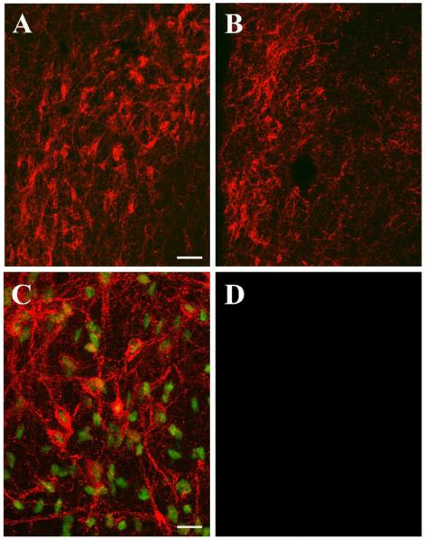
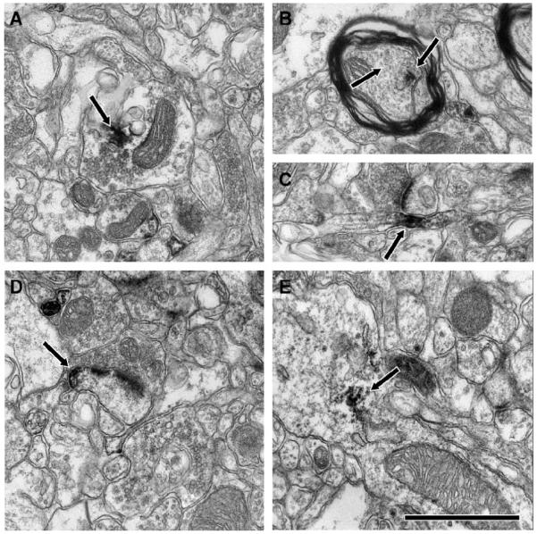
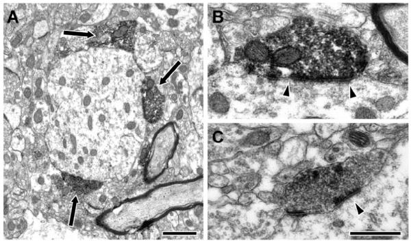
The transcription factor myocyte enhancer factor 2 (MEF2) is expressed throughout the central nervous system, where four MEF2 isoforms play important roles in neuronal survival and differentiation and in synapse formation and maintenance. It is therefore somewhat surprising that there is a lack of detailed information on the localization of MEF2 isoforms in the mammalian brain. We have analyzed the regional, cellular, and subcellular expression of MEF2A and MEF2D in the rodent brain. These two MEF2 isoforms were co-expressed in virtually all neurons in the cortex and the striatum, but were not detected in astrocytes. MEF2A and MEF2D were localized to the nuclei of neurons in many forebrain areas, consistent with their roles as transcriptional regulators. However, in several subcortical sites we observed extensive cytoplasmic expression of MEF2A but not MEF2D. MEF2A was particularly enriched in processes of neurons in the lateral septum and bed nucleus of the stria terminalis, as well as in several other limbic sites, including the central amygdala and paraventricular nuclei of the hypothalamus and thalamus. Ultrastructural examination similarly revealed MEF2A-ir in axons and dendrites as well as MEF2A-ir nuclei in the lateral septum and bed nucleus of the stria terminalis neurons. This study demonstrates for the first time extensive cytoplasmic localization of a MEF2 transcription factor in the mammalian brain in vivo. The extranuclear localization of MEF2A suggests novel roles for MEF2A in specific neuronal populations.
Figures
Similar articles
-
Myocyte enhancer factor 2A and 2D undergo phosphorylation and caspase-mediated degradation during apoptosis of rat cerebellar granule neurons.J Neurosci. 2001 Sep 1;21(17):6544-52. doi: 10.1523/JNEUROSCI.21-17-06544.2001. J Neurosci. 2001. PMID: 11517243 Free PMC article.
-
Regulation of the human GLUT4 gene promoter: interaction between a transcriptional activator and myocyte enhancer factor 2A.Proc Natl Acad Sci U S A. 2003 Dec 9;100(25):14725-30. doi: 10.1073/pnas.2432756100. Epub 2003 Nov 20. Proc Natl Acad Sci U S A. 2003. PMID: 14630949 Free PMC article.
-
Myocyte enhancer factor 2A is transcriptionally autoregulated.J Biol Chem. 2008 Apr 18;283(16):10318-29. doi: 10.1074/jbc.M707623200. Epub 2007 Dec 10. J Biol Chem. 2008. PMID: 18073218 Free PMC article.
-
Myocyte Enhancer Factor 2A Plays a Central Role in the Regulatory Networks of Cellular Physiopathology.Aging Dis. 2023 Apr 1;14(2):331-349. doi: 10.14336/AD.2022.0825. eCollection 2023 Apr 1. Aging Dis. 2023. PMID: 37008050 Free PMC article. Review.
-
The Molecular and Biological Function of MEF2D in Leukemia.Adv Exp Med Biol. 2024;1459:379-403. doi: 10.1007/978-3-031-62731-6_17. Adv Exp Med Biol. 2024. PMID: 39017853 Review.
Cited by
-
Measuring Nonapoptotic Caspase Activity with a Transgenic Reporter in Mice.eNeuro. 2022 Oct 4;9(5):ENEURO.0147-21.2022. doi: 10.1523/ENEURO.0147-21.2022. Print 2022 Sep-Oct. eNeuro. 2022. PMID: 36635920 Free PMC article.
-
Spine growth in the anterior cingulate cortex is necessary for the consolidation of contextual fear memory.Proc Natl Acad Sci U S A. 2011 May 17;108(20):8456-60. doi: 10.1073/pnas.1016275108. Epub 2011 Apr 29. Proc Natl Acad Sci U S A. 2011. PMID: 21531906 Free PMC article.
-
MEF2 negatively regulates learning-induced structural plasticity and memory formation.Nat Neurosci. 2012 Sep;15(9):1255-64. doi: 10.1038/nn.3189. Epub 2012 Aug 12. Nat Neurosci. 2012. PMID: 22885849
-
Inhibition of myocyte-specific enhancer factor 2A improved diabetic cardiac fibrosis partially by regulating endothelial-to-mesenchymal transition.Oncotarget. 2016 May 24;7(21):31053-66. doi: 10.18632/oncotarget.8842. Oncotarget. 2016. PMID: 27105518 Free PMC article.
References
-
- Black BL, Olson EN. Transcriptional Control of Muscle development by Myocyte Enhancer Factor-2 (MEF2) Proteins. Annu. Rev. Cell. Dev. Biol. 1998;14:167–196. - PubMed
-
- Cox DM, Du M, Marback M, Yang ECC, Chan J, Siu KWM, McDermott JC. Phosphorylation Motifs Regulating the Stability and Function of Myocyte Enhancer Factor 2A. J. Biol. Chem. 2003;278:15297–15303. - PubMed
Publication types
MeSH terms
Substances
Grants and funding
LinkOut - more resources
Full Text Sources
