. 2009 May 29;389(1):146-56.
doi: 10.1016/j.jmb.2009.04.005. Epub 2009 Apr 9.

U2504 determines the species specificity of the A-site cleft antibiotics: the structures of tiamulin, homoharringtonine, and bruceantin bound to the ribosome

Affiliations

U2504 determines the species specificity of the A-site cleft antibiotics: the structures of tiamulin, homoharringtonine, and bruceantin bound to the ribosome

Güliz Gürel et al. J Mol Biol. .

Abstract

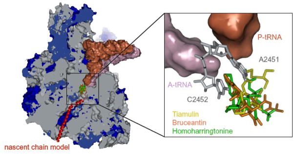
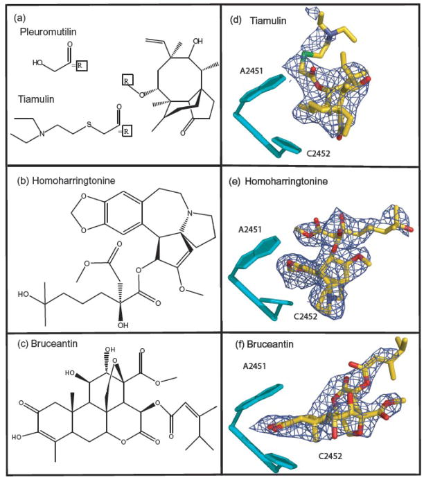
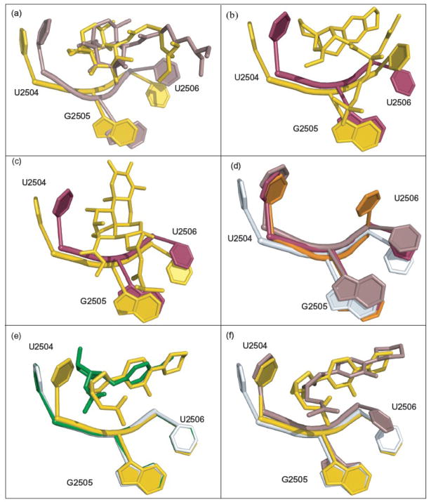
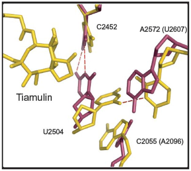
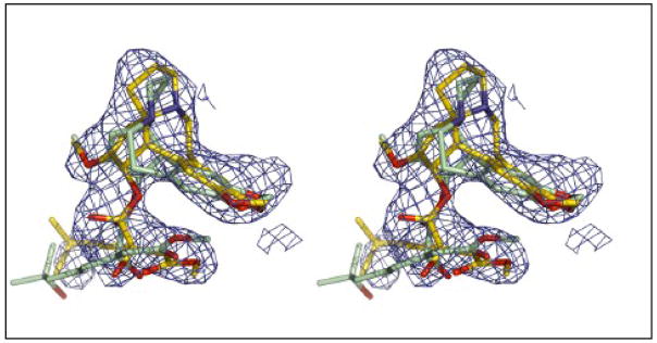
Structures have been obtained for the complexes that tiamulin, homoharringtonine, and bruceantin form with the large ribosomal subunit of Haloarcula marismortui at resolutions ranging from 2.65 to 3.2 A. They show that all these inhibitors block protein synthesis by competing with the amino acid side chains of incoming aminoacyl-tRNAs for binding in the A-site cleft in the peptidyl-transferase center, which is universally conserved. In addition, these structures support the hypothesis that the species specificity exhibited by the A-site cleft inhibitors is determined by the interactions they make, or fail to make, with a single nucleotide, U2504 (Escherichia coli). In the ribosome, the position of U2504 is controlled by its interactions with neighboring nucleotides, whose identities vary among kingdoms.


Figures

Figure 1
Figure 1
Cross-sectional view of the large ribosomal subunit. The subunit has been cut open to expose the peptidyl transferase center and the polypeptide exit tunnel. Ribosomal RNA is grey and proteins are blue. A P-site-bound tRNA (brown) is shown with an A-site-bound tRNA (purple) just visible behind it, and a nascent peptide chain (red) shown schematically extending down the exit tunnel. The site where the antibiotics of interest bind to the ribosome is also shown. The inset provides an enlarged view of the drug-binding site. tRNAs are shown as surfaces, color-coded as described above. The two nucleotides that define the A-site cleft are shown in gray, and the three drugs discussed here are shown superimposed on each other (tiamulin(yellow), homoharringtonine(green) and bruceantin(orange)).
Figure 2
Figure 2
The chemical structures of tiamulin, homoharringtonine and bruceantin, and the corresponding difference electron density. Panels (a), (b), and (c) show the chemical structures of these inhibitors. Panels (d), (e), and (f) show the feature in the appropriate (Fo-Fo) difference electron density map that were assigned to the three drugs with the structures of the drugs superimposed on them. Difference electron density maps were contoured at 3σ. Residues forming the A-site cleft are shown in cyan, and 23S rRNA bases are numbered to correspond with the 23S rRNA of E. coli.
Figure 3
Figure 3
Drug interactions with the ribosome, and the conformational changes associated with drug binding. Panels (a), (c), and (e) show how tiamulin, homoharringtonine, and bruceantin interact with the ribosome. Drugs are shown with C gold, N blue, O red, and S green. Water molecules are shown as red balls and hydrogen bonds are shown as red dashes. Panels (b), (d), and (f) show the conformational changes associated with drug binding. The drugs are color coded as just described. The drug-bound conformation of surrounding bases is shown in cyan, superimposed on the apo- conformation (gray).
Figure 4
Figure 4
Comparison of the interactions of tiamulin and homoharringtonine with archaeal and eubacterial ribosomes. (a) The structure of the complex tiamulin (gold) forms with the large ribosomal subunit from H. ma (gold) is shown superimposed on the structure of tiamulin (beige) bound to the large ribosomal subunit from D. radiodurans (beige) (PDB# 1XBP). (b) The structure of homoharringtonine (gold) bound to the H. ma large ribosomal subunit (gold) is shown superimposed on the structure of the E. coli large ribosomal subunit (PDB# 2AW4, magenta,). (c) The structure of bruceantine (gold) bound to H. ma large ribosomal subunit (gold) is shown superimposed on the structure of the E. coli large ribosomal subunit (PDB# 2AW4, magenta,). (d) The apo structures of H. ma (PDB# 1S72, gray,), T. thermophilus (PDB# 2J03, orange,), E. coli (PDB# 2AW4, magenta) and D. radiodurans (PDB# 1NKW, beige,). (e) The apo (PDB# 1S72, gray), anisomycin- bound (PDB# 1K73, green,) and linezolid-bound (PDB# 3CPW, gold,) structures of the H. marismortui large ribosomal subunit are superimposed. Anisomycin is green, and linezolid is gold. (f) A superposition of the apo structure of H. ma (PDB# 1S72, gray), the structure of the complex the H. ma ribosome forms with linezolid (PDB# 3CPW, gold), and the structure of linezolid bound to the D. radiodurans large ribosomal subunit (PDB# 3DLL, beige,). All superpositions in (a), (b), (c),(d), (e) and (f) were done using PTC phosphorus atom positions.
Figure 5
Figure 5
Interactions that control the conformation of U2504(2539) in H. ma and E. coli. The H. ma structure shown is the tiamulin-ribosome complex structure described here, and the E. coli structure is PDB# 2AW4. The two have been superimposed using phosphorus atom positions in the PTC. Tiamulin and the H. ma ribosome are shown in gold and the E. coli ribosome is in magenta.
Figure 6
Figure 6
Choosing the correct homoharringtonine enantiomer. A stereo pair is provided that compares the fit of the refined homoharringtonine structure (gold), structure to the electron density for that drug with the fit obtained for the homoharringtonine structure obtained from Cambridge Structural Database (green), which is its enantiomer. The electron density shown is Fo-Fo difference electron density contoured at 3σ.

References

    1. Hansen JL, Moore PB, Steitz TA. Structures of five antibiotics bound at the peptidyl transferase center of the large ribosomal subunit. J Mol Biol. 2003;330:1061–1075. - PubMed
    1. Wilson DN, Harms JM, Nierhaus KH, Schlunzen F, Fucini P. Species-specific antibiotic-ribosome interactions: implications for drug development. Biol Chem. 2005;386:1239–1252. - PubMed
    1. Vazquez D. Molecular Biology Biochemistry and Biopyhsics. Springer Verlag; New York: 1979. Inhibitors of Protein Biosynthesis. - PubMed
    1. Drews J, Georgopoulos A, Laber G, Schutze E, Unger J. Antimicrobial Activities of 81.723 Hfu, a New Pleuromutilin Derivative. Antimicrob Agents Chemother. 1975;7:507–516. - PMC - PubMed
    1. Kavanagh F, Hervey A, Robbins JW. Antibiotic Substances from Basidiomycetes. VIII Pleurotus Multilus (Fr.) Sacc and Pleurotus Passeckerianus Pilat. Proc Natl Acad Sci USA. 1951;37:570–574. - PMC - PubMed

Publication types