Review
. 2009 Oct;54(5):570-4.
doi: 10.1016/j.toxicon.2009.01.040. Epub 2009 Apr 9.

Association of botulinum neurotoxins with synaptic vesicle protein complexes

Affiliations
Review

Association of botulinum neurotoxins with synaptic vesicle protein complexes

Michael R Baldwin et al. Toxicon. 2009 Oct.

Abstract

Botulinum neurotoxins (BoNTs) elicit flaccid paralysis by cleaving SNARE proteins within peripheral neurons. BoNTs are classified into seven serotypes, termed A-G, based on antibody cross-neutralization. Clostridia produce BoNTs as single-chain toxins that are cleaved into a di-chain protein that comprises an N-terminal zinc metalloprotease domain that is linked by a disulfide bond to the C-terminal translocation/receptor-binding domain. BoNT/A and BoNT/B utilize synaptic vesicle protein 2 (SV2) and synaptotagmin, respectively, as receptors for entry into neurons. Using affinity chromatography, BoNT/A and BoNT/B were found to bind a synaptic vesicle protein complex in CHAPS extracts of synaptic vesicles. Mass spectroscopy identified synaptic vesicle protein 2, synaptotagmin I, synaptophysin, vesicle-associated membrane protein 2, and the vacuolar ATPase-proton pump as components of the BoNT-synaptic vesicle protein complex. BoNT/A and BoNT/B possessed unique density-gradient profiles when bound to synaptic vesicle protein complexes. The identification of BoNT/A and BoNT/B bound to synaptic vesicle protein complexes provides insight into the interactions of BoNT and neuronal receptors.


Figures
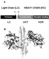
Figure 1
Figure 1. Structure-function properties of the botulinum neurotoxins
(A) Ribbon diagram of BoNT/A (pdb: 3bta). (B) Linear diagram of the di-chain activated BoNT molecule consisting of a light (LC) and heavy chain (HC) bound by a single disulfide bond. HC is divided into an N-terminal translocation domain (HCT) and a C-terminal receptor binding domain (HCR).
Figure 2
Figure 2. Model for binding and entry of BoNTs at the Neuromuscular junction
BoNT/A associates with the presynaptic membrane of α-motor neurons through interactions with oligosaccharides such as ganglioside GT1b (1). Calcium influx stimulates synaptic vesicle membrane fusion (2). BoNT/A interacts with the exposed receptor complex, stimulating synaptic vesicle recycling (3). Recycled synaptic vesicles are acidified through the activity of the v-ATPase (4). Vesicle acidification drives the translocation of the BoNT/A light chain (LC/A) into the cytosol (5) completing the cycle (6).
Figure 3
Figure 3. Protocol for the identification of BoNT-HCR-binding proteins
Rat brain cortex are homogenized and subjected to differential centrifugation. Synaptic vesicles are lysed and extracted with either CHAPS or Triton-X100. A 3-FLAG epitope IP is performed to isolate HCR binding proteins. 3FLAG peptide is added to the IP to release #-FLAG-HCR form the bead along with HCR biding proteins, which are identified by MALDI-MS or Western blotting.
Figure 4
Figure 4. Model for binding of BoNTs to the synaptic vesicle protein complex
Synaptic vesicle proteins forms a large protein complex that includes synaptic vesicle protein 2 (SV2), synaptotagmin I, synaptophysin (Syp), vesicle-associated membrane protein 2 (VAMP 2) and the vacuolar proton pump (v-ATPase). The synaptic receptor complex is stable following solubilization with CHAPS and HCR/A binds to SV2 and HCR/B binds synaptotagmin I within the complex.

Similar articles

Cited by

References

    1. Baldwin MR, Barbieri JT. Association of botulinum neurotoxin serotypes a and B with synaptic vesicle protein complexes. Biochemistry. 2007;46:3200–3210. - PubMed
    1. Belizaire R, Komanduri C, Wooten K, Chen M, Thaller C, Janz R. Characterization of synaptogyrin 3 as a new synaptic vesicle protein. J Comp Neurol. 2004;470:266–281. - PubMed
    1. Bennett MK, Calakos N, Kreiner T, Scheller RH. Synaptic vesicle membrane proteins interact to form a multimeric complex. J Cell Biol. 1992;116:761–775. - PMC - PubMed
    1. Blasi J, Chapman ER, Link E, Binz T, Yamasaki S, De Camilli P, Sudhof TC, Niemann H, Jahn R. Botulinum neurotoxin A selectively cleaves the synaptic protein SNAP-25. Nature. 1993a;365:160–163. - PubMed
    1. Blasi J, Chapman ER, Yamasaki S, Binz T, Niemann H, Jahn R. Botulinum neurotoxin C1 blocks neurotransmitter release by means of cleaving HPC-1/syntaxin. Embo J. 1993b;12:4821–4828. - PMC - PubMed

Publication types

MeSH terms