. 2009 Aug;1791(8):746-56.
doi: 10.1016/j.bbalip.2009.03.016. Epub 2009 Apr 9.

Serine palmitoyltransferase subunit 1 is present in the endoplasmic reticulum, nucleus and focal adhesions, and functions in cell morphology

Affiliations

Serine palmitoyltransferase subunit 1 is present in the endoplasmic reticulum, nucleus and focal adhesions, and functions in cell morphology

Jia Wei et al. Biochim Biophys Acta. 2009 Aug.

Abstract

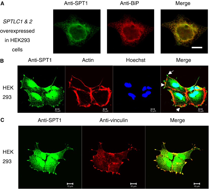
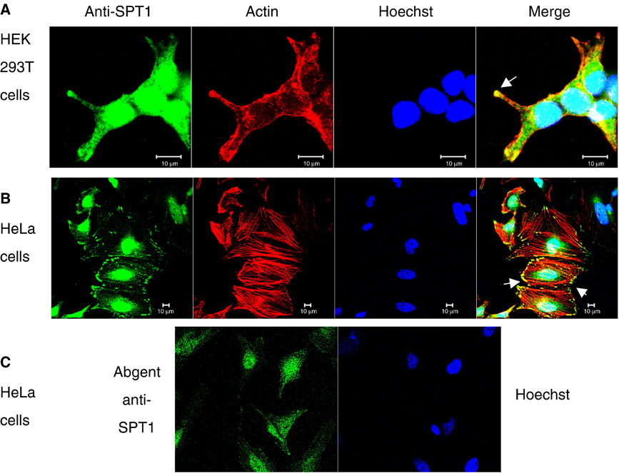
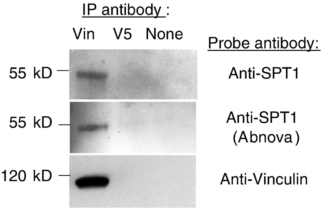
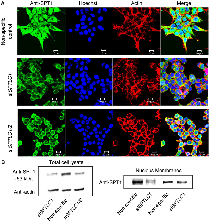
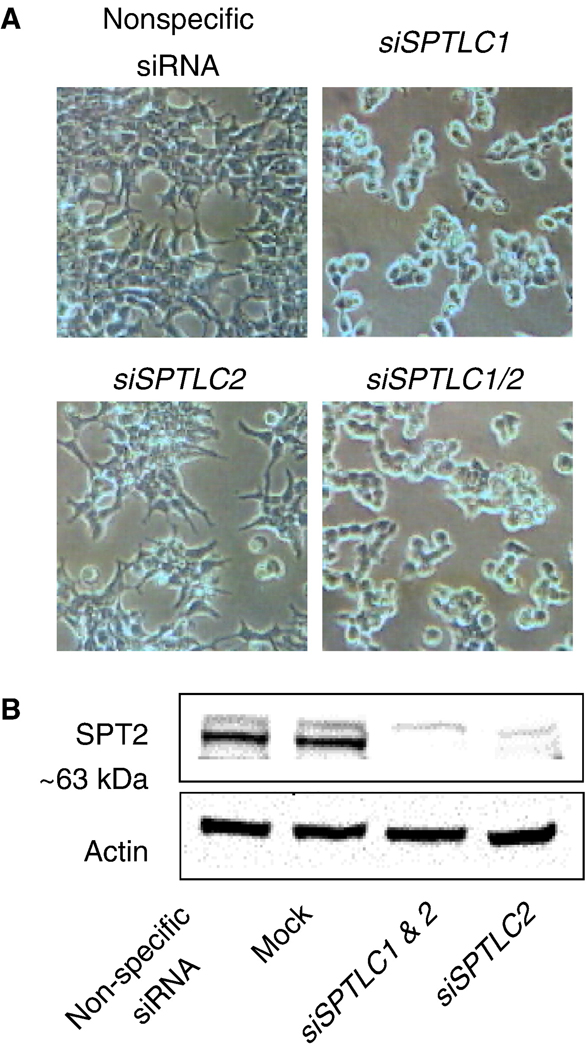
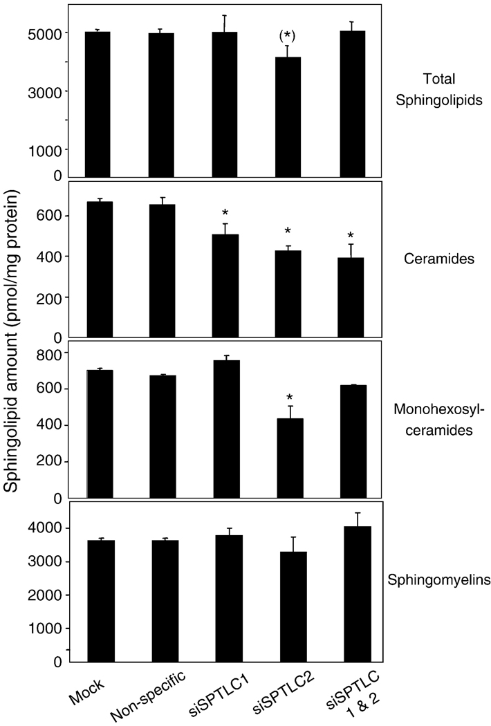
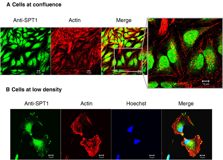
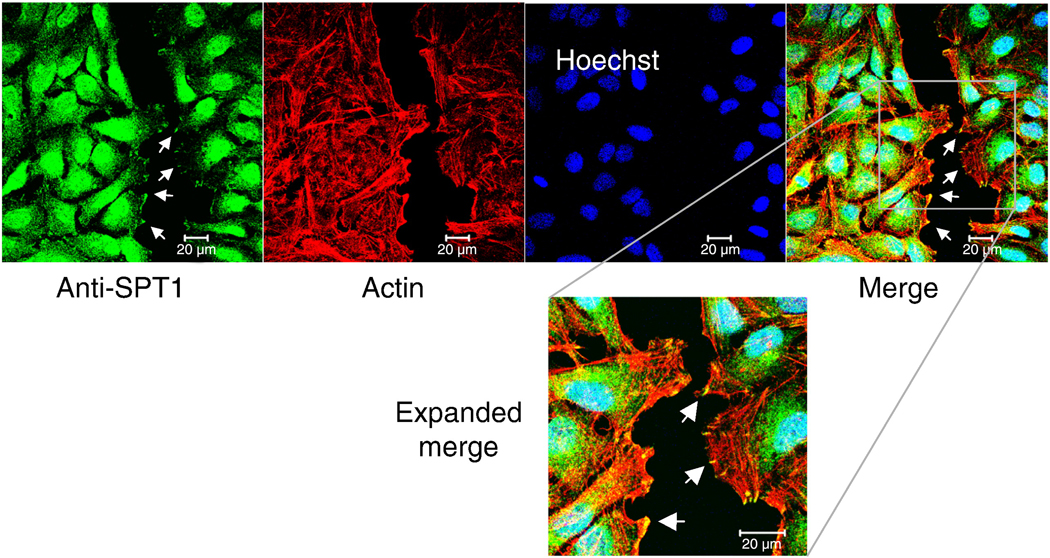
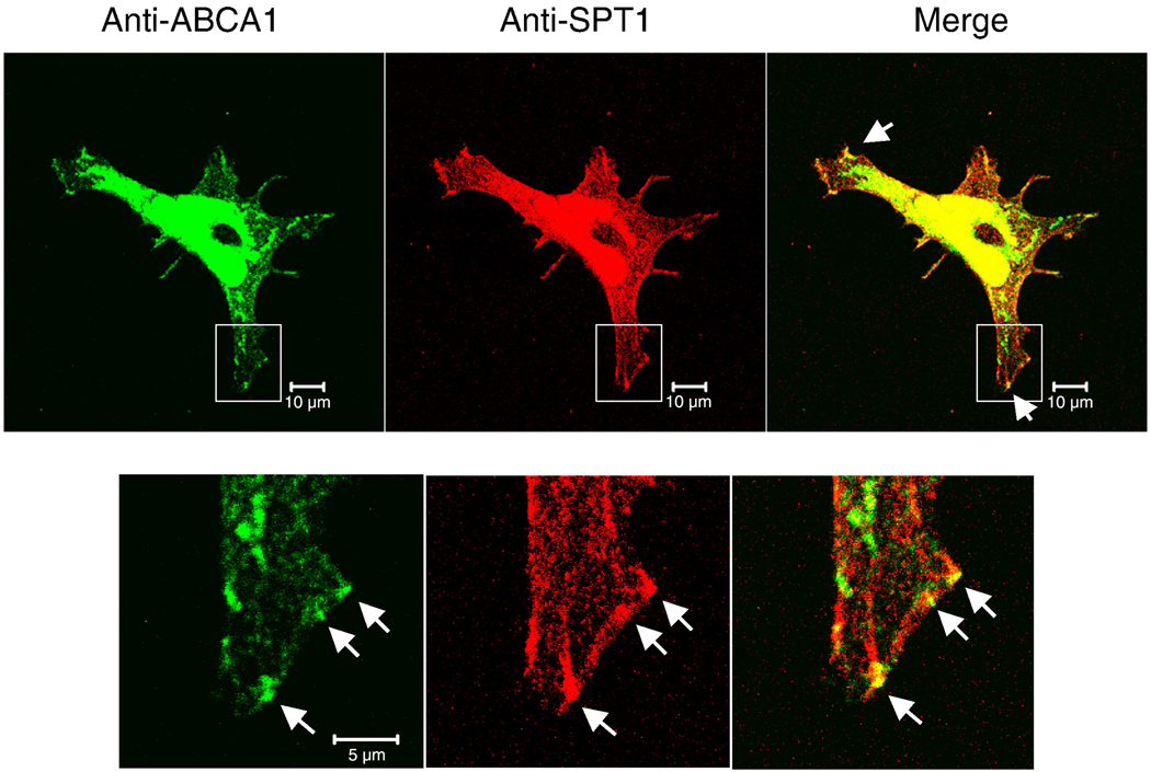
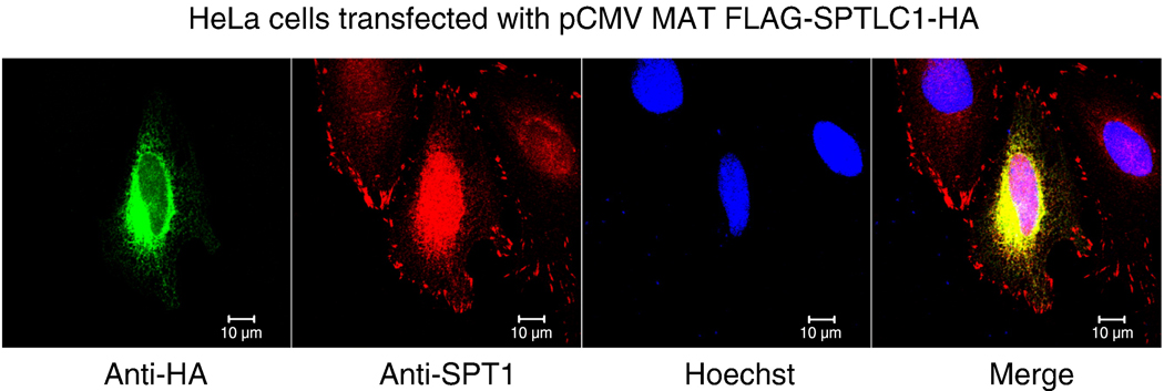
Serine palmitoyltransferase (SPT) has been localized to the endoplasmic reticulum (ER) by subcellular fractionation and enzymatic assays, and fluorescence microscopy of epitope-tagged SPT; however, our studies have suggested that SPT subunit 1 might be present also in focal adhesions and the nucleus. These additional locations have been confirmed by confocal microscopy using HEK293 and HeLa cells, and for focal adhesions by the demonstration that SPT1 co-immunoprecipitates with vinculin, a focal adhesion marker protein. The focal adhesion localization of SPT1 is associated with cell morphology, and possibly cell migration, because it is seen in most cells before they reach confluence but disappears when they become confluent, and is restored by a standard scratch-wound healing assay. Conversely, elimination of SPT1 using SPTLC1 siRNA causes cell rounding. Thus, in addition to its "traditional" localization in the ER for de novo sphingolipid biosynthesis, SPT1 is present in other cellular compartments, including focal adhesions where it is associated with cell morphology.


Figures

Fig. 1
Fig. 1
Intracellular localization of SPT1 in SPTLC1&SPTLC2 overexpressing HEK293 cells and native HEK293 cells by immunofluorescence staining and confocal microscopy. Cells were grown on glass coverslips for 24 h before collecting for immunostaining. Rabbit anti-SPT1 antibody was used as primary labeling and Alexa Fluor 488 anti-rabbit antibody (green) as secondary labeling. (A) Localization of SPT1 in HEK293/SPTLC1/2 stable cells. SPT1 protein was stained in green and the ER marker protein, Bip, was labeled with Alexa Flour 568 (red). (B) Localization of endogenous SPT1 proteins in HEK293 cells. SPT1 was labeled in green. Actin was labeled by rhodamin phalloidin (red) and the nucleus was by Hoescht 33342 (blue). The arrows point to the focal adhesion localization of SPT1 at the tips of actin stress fibers. (C) Co-localization of SPT1 and vinculin in HEK293 cells. Mouse anti-vinculin antibody was used as a primary labeling followed by Alexa Flour 568 anti-mouse antibody (red). The images were taken by Zeiss confocal microscope. Bar, 10 µm.
Fig. 2
Fig. 2
SPT1 staining in HEK293T cells and HeLa cells. Confocal image of SPT1 in HEK293T cells (A) and HeLa cells (B) by rabbit anti-SPT1 antibody labeled by Alexa Fluor 488 anti-rabbit antibody (green), and (C) for HeLa cells using another rabbit anti-SPT1 antibody (catalog # AP2534b from Abgent, San Diego, CA). Actin was labeled by rhodamine phalloidin (red) and the nucleus was by Hoescht 33342 (blue).
Fig. 3
Fig. 3
Coimmunoprecipitation of SPT1 by an anti-vinculin antibody. A HEK293 cell lysate was treated with a mouse anti-vinculin antibody (left lane), an antibody against a protein not found in mammalian cells (mouse anti-V5) (middle lane), or no antibody (right lane) followed by recovery of the capture antibodies using protein G sepharose beads, then analysis of the precipitates by Western blotting (right lane). The immunoprecipitates were applied for SDS-PAGE and immunoblotted with the rabbit anti-SPT1 antibody used for the confocal imaging of SPT1 with focal adhesions (top panel), a commercial rabbit SPT1 antibody (Abnova, middle panel) and an anti-vinculin antibody (bottom panel).
Fig. 4
Fig. 4
Confocal imaging of SPT1 in HEK293T cells transfected with siRNA to silence SPTLC1. (A) HEK293T cells grown on collagen coated coverslips were transfected with non-specific siRNA, siSPTLC1 and siSPTLC1/siSPTLC2 separately for 72 h and then fixed for immunofluorescent staining using anti-SPT1 antibody and Alexa Fluor 488 anti-rabbit antibody (green) as well as for actin fibers (phalloidin) and nuclei (Hoechst 3342) (Bar, 10 µm); or (B) analyzed by western blotting for SPT1 normalized by actin. The right immunoblot is for the nuclear and membrane fractions isolated from HEK293T cells transfected with either non-specific siRNA or siSPTLC1 as described in experimental procedures.
Fig. 5
Fig. 5
Morphology of HEK293T cells after transfection with control and SPTLC siRNAs. (A) Phase contrast images of HEK293T cells transfected with non-specific siRNA, siSPTLC1, siSPTLC2 and siSPTLC1/siSPTLC2 separately. (B) The western blots that show that SPT2 protein has been suppressed by siSPTLC2 alone and in combination with siSPTLC1, with normalization using actin.
Fig. 6
Fig. 6
Amounts of ceramide, monohesoxylceramides and sphingomyelins in HEK293T cells after transfection with control vectors or siSPTLC1 and/or siSPTLC2. HEK293T cells were transfected with non-specific siRNA, siSPTLC1, siSPTLC2 and siSPTLC1/SPTLC2 separately for 72 h and then collected for lipid extraction and analysis by LC ESI MS/MS. The amounts are given as the mean ± SD for analysis of 2 dishes.
Fig. 7
Fig. 7
Focal adhesion staining of SPT1 in low density cells and confluent cells. (A) Confocal fluorescence imaging of SPT1 in confluent HeLa cells. HeLa cells were grown on collagen coated coverslips until confluent and then fixed for immunofluorescent stain with anti-SPT1 antibody followed by Alexa Fluor 488 anti-rabbit antibody (note enlarged image). (B) SPT1 in low density cells. Cells were cultured at low density before applied for immunofluorescence staining. The two cells shown here appear to be at a late stage of mitosis. Actin fibers and nuclei were labeled by rhodamine phalloidin and Hoechst 3342 separately. Bar, 10 µm.
Fig. 8
Fig. 8
Focal adhesion staining of SPT1 during the scratch wound-healing assay. HeLa cells were grown to confluent on collagen coated glass coverslips, scratched by a pipet tip, and processed for immunofluorescence staining using anti-SPT1 antibody after 3 h. The bottom image is an enlargement of the shown part of the merged image. The arrows point to some of the focal adhesions. Bar, 10 µm.
Fig. 9
Fig. 9
Localization of SPT1 and ABCA1 in HEK cells. HEK293 cells grown on collagen coated coverslips were stained with rabbit anti-SPT1 antibody and mouse anti-ABCA1 antibody followed by Alexa Fluor 488 anti-rabbit antibody (green) and Alexa Flour 568 anti-mouse antibody (red). The enlarged images show the localization similarity of the two proteins at the leading edge of the cell. Upper panel: bar, 10 µm; lower panel: bar, 5 µm.
Fig. 10
Fig. 10
Localization of SPT1 in SPT1-overexpressing HeLa cells. HeLa cells were transfected with pCMV MAT FLAG-SPTLC1-HA and then stained by mouse anti-HA and rabbit anti-SPT1 antibody followed by anti-mouse Alexa Flour 488 (green) and anti-rabbit Alexa Flour 568 (red). Bar, 10 µm.

Similar articles

Cited by

References

    1. Alvarez SE, Milstien S, Spiegel S. Autocrine and paracrine roles of sphingosine-1-phosphate. Trends Endocrinol. Metab. 2007;18:300–307. - PubMed
    1. Hannun YA, Obeid LM. Principles of bioactive lipid signalling: lessons from sphingolipids. Nat. Rev. 2008;9:139–150. - PubMed
    1. Merrill AH, Jr, Wang MD, Park M, Sullards MC. (Glyco)sphingolipidology: an amazing challenge and opportunity for systems biology. Trends Biochem. Sci. 2007;32:457–468. - PubMed
    1. Spiegel S, Milstien S. Sphingosine-1-phosphate: an enigmatic signalling lipid. Nat. Rev. 2003;4:397–407. - PubMed
    1. Todeschini AR, Dos Santos JN, Handa K, Hakomori SI. Ganglioside GM2/GM3 complex affixed on silica nanospheres strongly inhibits cell motility through CD82/cMet-mediated pathway. Proc. Natl. Acad. Sci. U S A. 2008;105:1925–1930. - PMC - PubMed

Publication types

MeSH terms