Review
. 2009 Jun;19(3):267-76.
doi: 10.1016/j.sbi.2009.03.005. Epub 2009 Apr 9.

Toward a structural understanding of IRES RNA function

Affiliations
Review

Toward a structural understanding of IRES RNA function

Megan E Filbin et al. Curr Opin Struct Biol. 2009 Jun.

Abstract

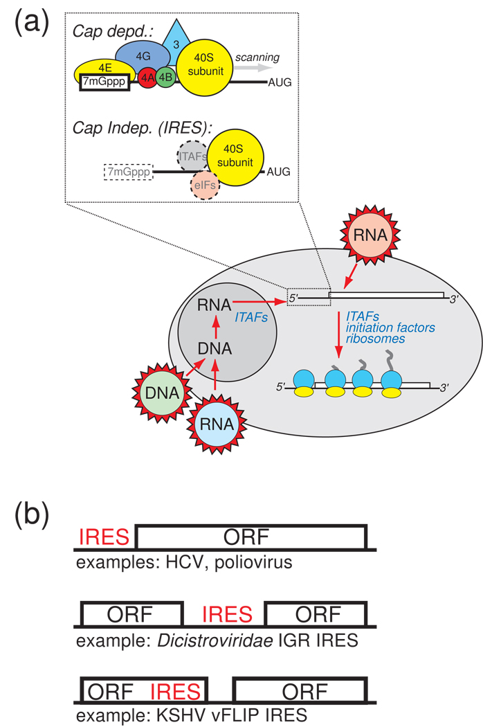
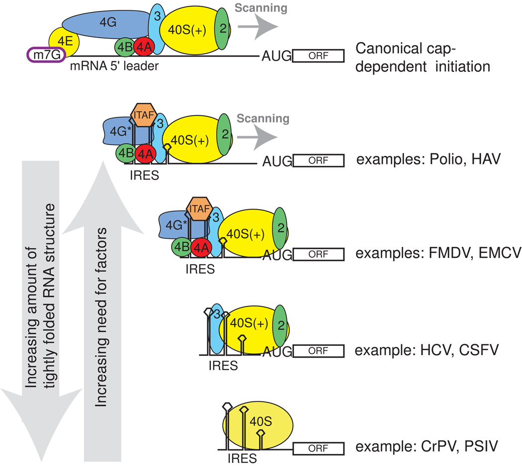
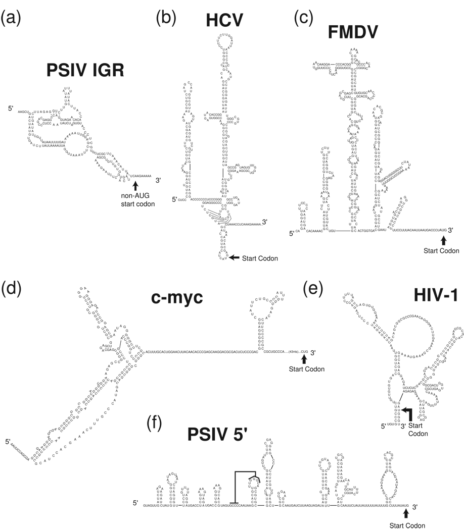
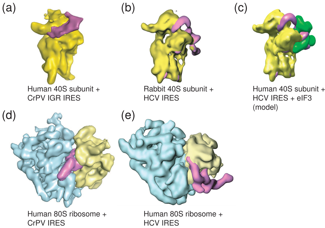
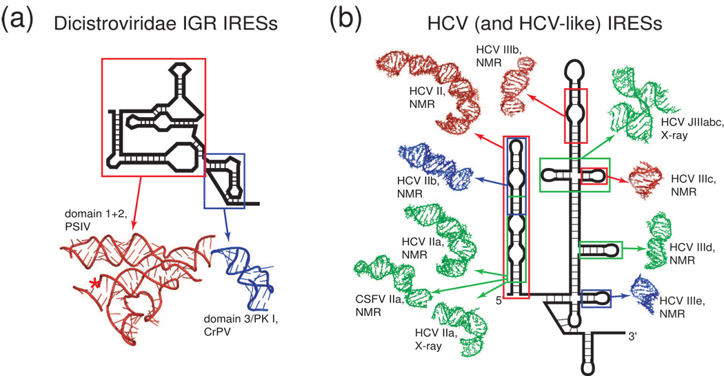
Protein synthesis of an RNA template can start by two different known mechanisms: cap-dependent translation initiation and cap-independent translation initiation. The latter is driven by RNA sequences called internal ribosome entry sites (IRESs) that are found in both viral RNAs and cellular mRNAs. The diverse mechanisms used by IRESs are reflected in their structural diversity, and this structural diversity challenges us to develop a cohesive model linking IRES function to structure. With more direct structural information available for the viral IRESs, data suggest an inverse correlation between the degree to which an IRES RNA can form a stable structure on its own and the number of factors that it requires to function. Lessons learned from the viral IRESs may help understand the cellular IRESs, although more structural data are needed before any strong links can be made.


Figures

Figure 1
Figure 1. Cap- and IRES- dependent translation initiation
(a) At upper left is a cartoon illustrating a simplified mechanism for IRES-driven initiation compared to cap-dependent initiation. In cap-dependent translation, the 7-methyl guanosine cap is bound by eIF4E, and this leads to the binding of many other factors, recruitment of the ribosome, and scanning to the start codon. IRESs do not use the cap structure, although IRES-containing messages can be capped (dashed box), and they may or may not use canonical eIFs and ITAFs to recruit the ribosome to the message (dashed circles). The diagram below illustrates the sources of IRES RNAs in the cell. They can come from RNA viruses that introduce RNA directly into the cytoplasm (red), and thus never experience the nuclear environment. IRESs that do have a “nuclear history” include those from RNA viruses whose genetic information is reverse transcribed and integrated into the host’s genome (blue), DNA viruses (green), and cellular IRESs. The degree to which nuclear history plays a role in the binding of specific factors to certain IRESs, and the effect it may have on the structure of the IRES, is a very important question under exploration. (c) Some mRNA contexts in which IRESs are found. Most are found in the 5’ untranslated region of the mRNA or viral RNA (top), but some are found between open reading frames in intergenic regions (middle), and they can also reside within (or partially within) coding regions (bottom). Viral IRES examples are provided for each.
Figure 2
Figure 2. Examples of the diversity of viral IRES factor requirements
Canonical initiation requires the full complement of translation eIFs (top), while IRES initiation can use subsets of these factors as well as ITAFs (below). Shown are examples of some viral IRESs with the factors each requires. For simplicity, all the factors associated with the 40S subunit are not shown. As described in the text, we note a trend in which IRES RNAs with the most inherent stably folded structure (left arrow) are those that require the fewest factors, and as the IRES become less inherently structured, more ITAFs and eIFS are needed (right arrow). The degree to which this trend will prove predictive, or can be extended to cellular IRESs, is unknown.
Figure 3
Figure 3. Examples of viral and cellular IRES secondary structures
Experimentally tested secondary structures of several diverse viral and cellular IRES RNAs are shown. (a) Plautia stali intestine virus (PSIV) IGR IRES. (b) HCV IRES (c) FMDV IRES (d) c-myc IRES (e) Human immunodeficiency virus-1 (HIV-1) gag- IRES (f) PSIV 5' IRES, the black line indicates a proposed pseudoknot interaction. Note that these secondary structures may be revised as more information becomes available regarding differences between RNA made and folded in vitro versus that made in vivo, which folds co-transcriptionally.
Figure 4
Figure 4. Cryo-EM reconstructions of two different viral IRESs bound to the ribosome
To date, only the HCV IRES (and related) and the Dicistroviridae IRES RNAs have been shown to bind directly to ribosomal subunits, and these complexes have been studies by cryo-EM. (a) The Cricket paralysis virus (CrPV) IGR IRES (magenta) bound to the 40S subunit (yellow). (b) The HCV IRES (magenta) bound to a 40S subunit (yellow). These two IRES types occupy different sites on the subunit, but both induce a similar conformational change in the 40S subunit when they bind, suggesting some mechanistic convergence. (c) Model of the HCV IRES bound to the 40S subunit and eIF3 (green), built from several cryo-EM reconstructions. (d) The CrPV IGR IRES bound to an 80S ribosome, the view is rotated 90 degrees from the view in panel (a). Note that higher resolution reconstructions of this complex have been published, but to allow more direct comparison with the HCV IRES-bound ribosome, the middle resolution structure is shown. (e) The HCV IRES bound to an 80S ribosome. Again, the different binding modes of these two IRESs to the ribosome are obvious, as are differences in the overall folded architectures of the IRESs; the CrPV IGR IRES is more compact, while the HCV IRES is extended. Cryo-EM reconstructions of preiniation complexes containing other IRESes have not been reported.
Figure 5
Figure 5. IRES RNA domains whose structures have been solved by X-ray crystallography or NMR
The structures of only a handful of IRES RNA domains have been determined. (a) Secondary structure cartoon of a Dicistroviridae IGR IRES with ribbons diagrams of the two structural domains of this IRES that have been solved by X-ray crystallography. This is the only IRES for which a complete structural picture exists. (b) Secondary structure cartoon of the HCV IRES surrounded by structures of various domains (shown in various colors) from both it and the CSFV IRES that have been solved by NMR or crystallography [–31]. For both the HCV and the Dicistroviridae IGR IRESs, these structures have been combined with cryo-EM reconstructions to develop models for how the IRES RNAs interact with the ribosome (not shown).
Figure 6
Figure 6. Models for ribosome recruitment by IRES•eIF•ITAF complexes
Within complex “IRES RNPs,” does the ribosome contact only the bound eIFs, a combination of eIFs and the IRES RNA, a combination of eIFs and ITAFs, etc.? The answer to this question may vary depending on the IRES, and awaits more structural information. A few possibilities are diagrammed here. (a) The ITAF could stabilize the active conformation of the IRES RNA, which is bound by eIFs that interact with the ribosome, but without direct interaction between the IRES RNA and the ribosome. (b) The ITAF and eIFs could both interact directly with the ribosome, again with no direct IRES RNA-ribosome interactions. (c) The ITAF could stabilize the active conformation of the IRES RNAs, and both the IRES and eIFs could contact the ribosome. (d) The ITAF, the eIFs and the RNA could all directly contact the ribosome. Note that other combinations could occur and that these are not mutually exclusive possibilities.

Similar articles

Cited by

References

    1. Pestova T, Lorsch JR, Hellen CU. The Mechanism of Translation Initiation in Eukaryotes. In: Mathews MB, Sonenberg N, editors. Translational Control in Biology and Medicine. Hershey JWB: Cold Sring Harbor Laboratory Press; 2007. pp. 87–128.
    1. Doudna JA, Sarnow P. Translation initiation by viral internal ribosome entry sites. In: Mathews MB, Sonenberg N, editors. Translational Control in Biology and Medicine. Hershey JWB: Cold Spring Harbor Laboratory Press; 2007. pp. 129–153.
    1. Elroy-Stein O, Merrick WC. Translation initiation via cellular internal ribosome entry sites. In: Mathews MB, Sonenberg N, Hershey J, editors. Translational Control in Biology and Medicine. Hershey J: Cold Spring Harbor Laboratroy Press; 2007. pp. 155–172.
    1. Jackson RJ. Alternative mechanisms of initiating translation of mammalian mRNAs. Biochem Soc Trans. 2005;33:1231–1241. - PubMed
    1. Baird SD, Turcotte M, Korneluk RG, Holcik M. Searching for IRES. RNA. 2006;12:1755–1785. - PMC - PubMed

Publication types