. 2009 Apr 10;34(1):93-103.
doi: 10.1016/j.molcel.2009.03.008.

The fic domain: regulation of cell signaling by adenylylation

Affiliations

The fic domain: regulation of cell signaling by adenylylation

Carolyn A Worby et al. Mol Cell. .

Abstract

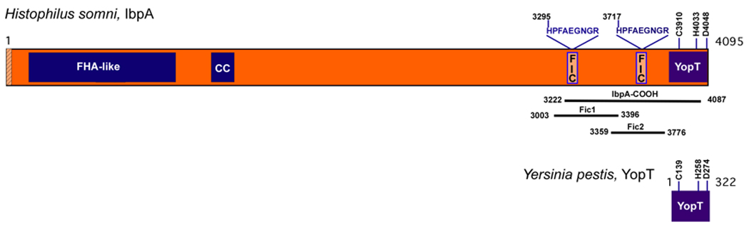
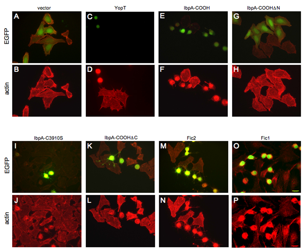
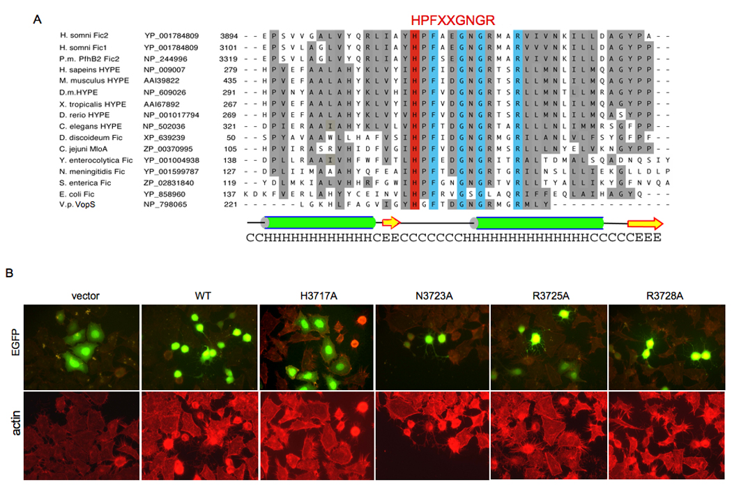
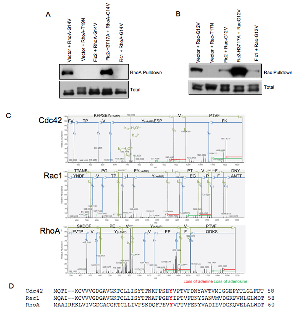
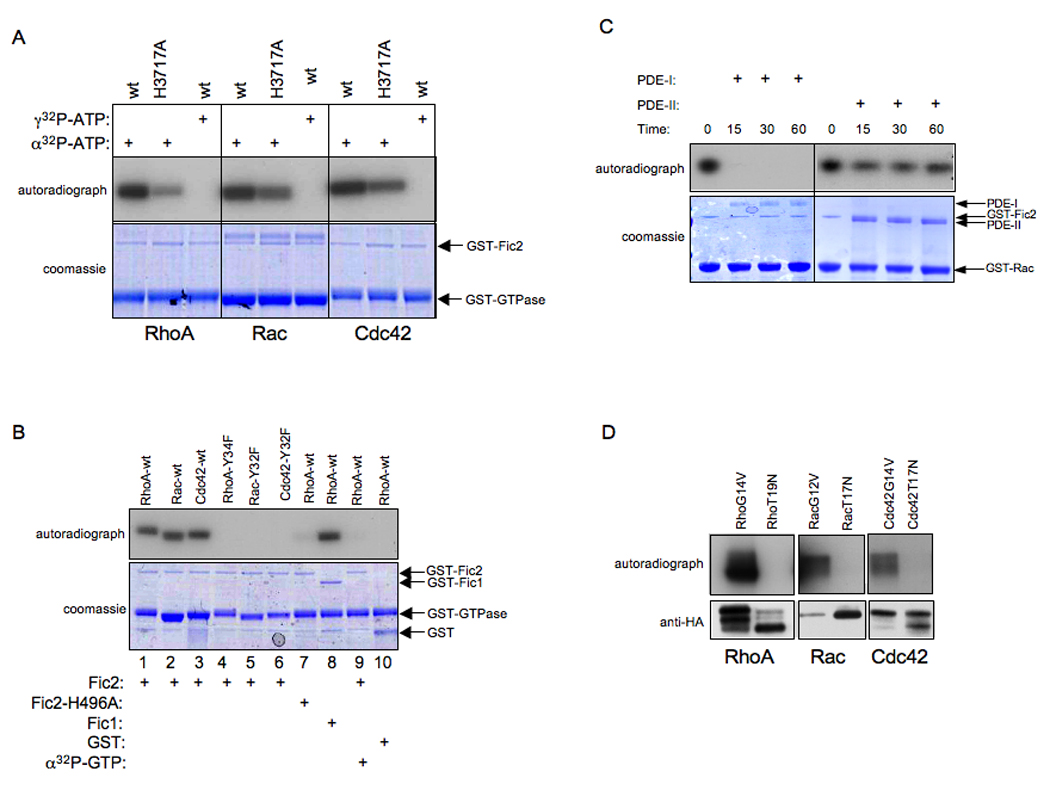
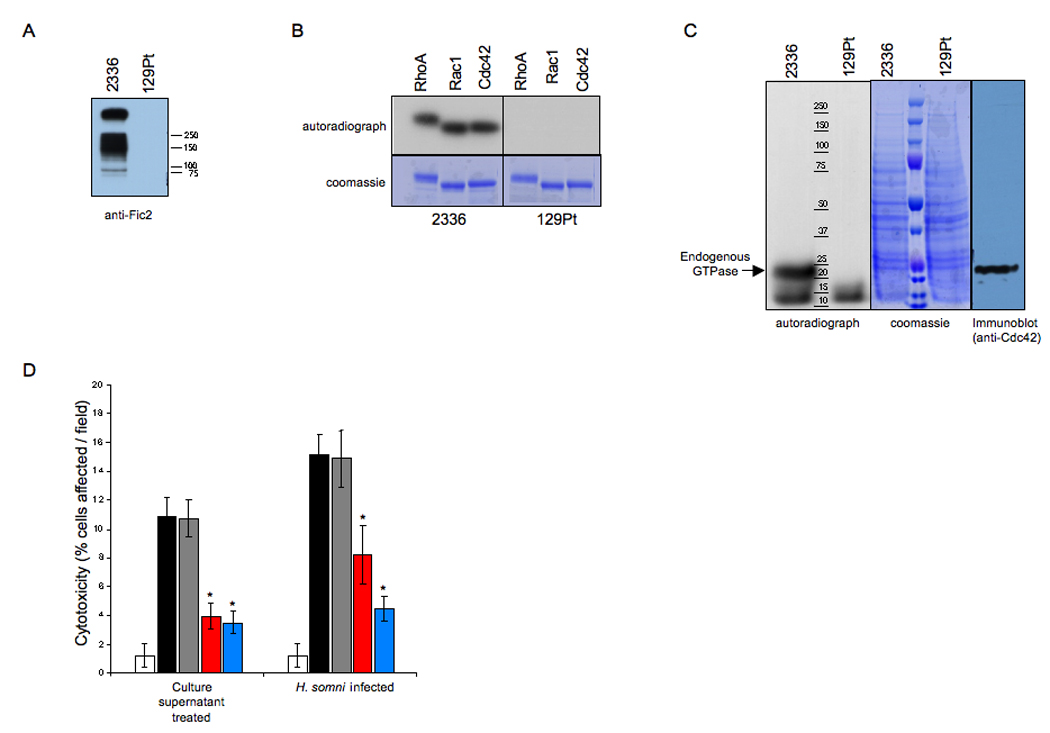
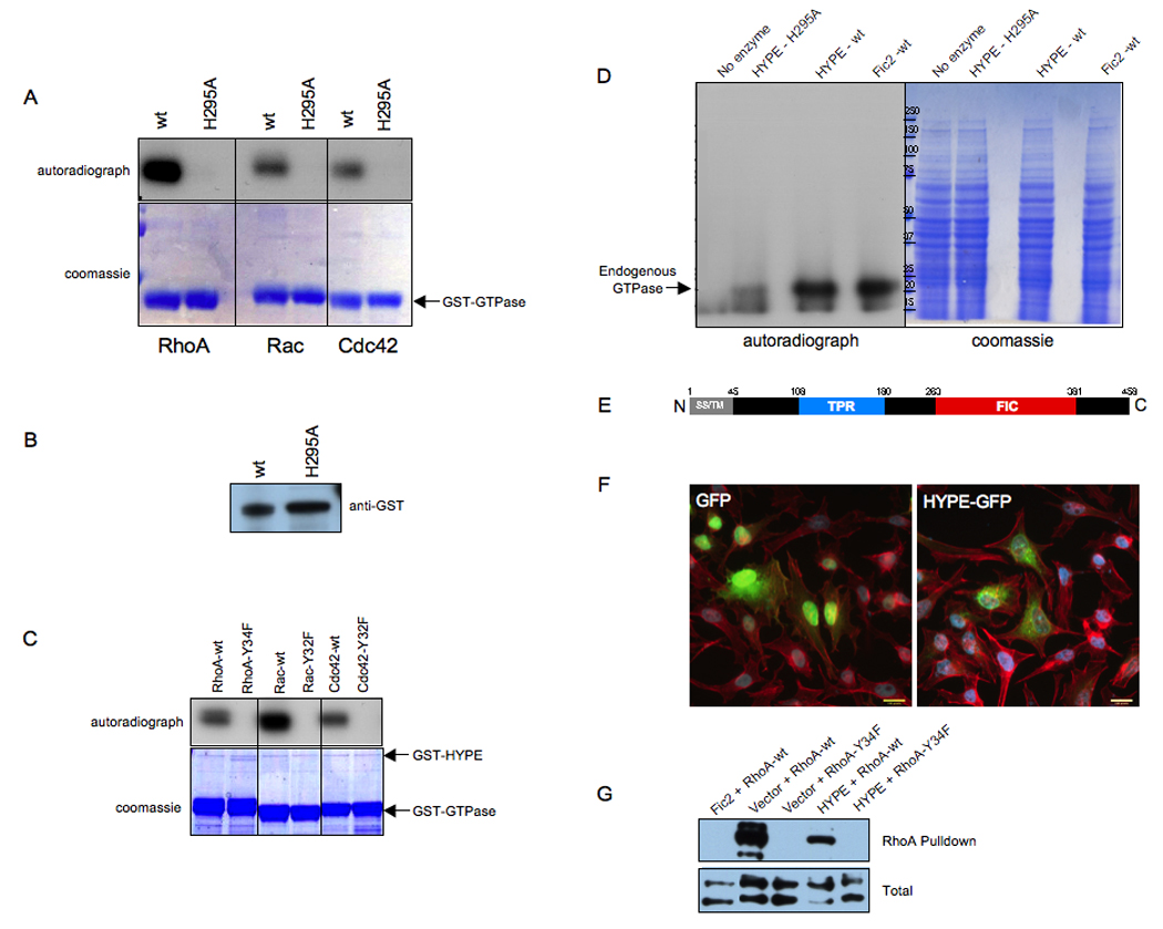
We show that the secreted antigen, IbpA, of the respiratory pathogen Histophilus somni induces cytotoxicity in mammalian cells via its Fic domains. Fic domains are defined by a core HPFxxGNGR motif and are conserved from bacteria to humans. We demonstrate that the Fic domains of IbpA catalyze a unique reversible adenylylation event that uses ATP to add an adenosine monophosphate (AMP) moiety to a conserved tyrosine residue in the switch I region of Rho GTPases. This modification requires the conserved histidine of the Fic core motif and renders Rho GTPases inactive. We further demonstrate that the only human protein containing a Fic domain, huntingtin yeast-interacting protein E (HYPE), also adenylylates Rho GTPases in vitro. Thus, we classify Fic domain-containing proteins as a class of enzymes that mediate bacterial pathogenesis as well as a previously unrecognized eukaryotic posttranslational modification that may regulate key signaling events.


Figures

Figure 1
Figure 1. Architecture of H. somni IbpA
IbpA contains an N-terminal secretion signal (hatched rectangle) followed by a filamentous hemagglutinin-like domain (FHA-like, blue box). Internal coiled-coils (CC, blue boxes) precede two Fic containing domains (FIC, light orange boxes). The YopT homology domain is present at the C-terminus (YopT, blue box) and the invariant catalytic C/H/D residues of IbpA and Y. pestis YopT are indicated. The invariant residues of IbpA’s Fic motif, HPFAEGNGR, are noted. The IbpA-COOH, Fic1 and Fic2 sequences are shown. All sequences are numbered from the initiator methionine for IbpA.
Figure 2
Figure 2. The Fic containing domains of IbpA are cytotoxic
Immunofluorescence microscopy was performed on HeLa cells transfected with the EGFP vector control and EGFP-YopT, EGFP-IbpA-COOH, or the designated EGFP-IbpA deletion constructs. Cell transfection, IbpA construct expression, and cell morphology of the indicated constructs were visualized by EGFP fluorescence (A, C, E, G, I, K, M, O). Actin filaments were visualized by rhodamine phalloidin staining (B, D, F, H, J, L, N,P). Expression of EGFP alone is not cytotoxic (A, B), while expression of both EGFP-YopT (C, D) and EGFP-IbpA-COOH (E, F) induces cytotoxicity. This cytotoxic activity was further narrowed down to the Fic2 (M, N) and Fic1 (O, P) domains of IbpA.
Figure 3
Figure 3. Identification of a Fic core motif
(A) Multiple sequence alignment of the Fic motifs. The Fic family was identified by BLAST searches and aligned by Clustal-W and Promals3D web programs (Pei et al., 2008), and manually adjusted using secondary structure predictions from PSIPRED (McGuffin et al., 2000). The two Fic containing domains of IbpA, Fic1 and Fic2 bear greatest sequence similarity to the two Fic containing domains of P. multocida, PfhB2, followed by mammalian HYPE. Selected Fic motifs displaying their diverse evolutionary conservation in descending order of similarity to IbpA’s Fic2 domain are shown. Listed from left to right are species, protein, accession number, and amino acids. Identities are boxed in blue. Similarities are shaded grey. The Fic HPFxxGNGR active site motif and the catalytic His residue are highlighted in red. The conserved predicted secondary structure for the Fic motif based on the prediction for Fic2 is diagramed; alpha-helices (H) are green; beta-strands (E) are yellow. The species listed are Histophilus somni, Pasteurella multocida (P.m.), Homo sapiens, Mus musculus, Drosophila melanogaster (D.m.), Xenopus tropicalis, Danio rerio, Caenorhabditis elegans, Dictyostelium discoideum, Campylobacter jejuni, Yersinia enterocolytica, Neisseria meningitidis, Salmonella enterica, Escherichia coli, and Vibrio parahemolyticus (V.p.). (B) Histidine 3717 is critical for Fic2 cytotoxicity. The invariant putative active site residues identified in the Fic family members were mutated in the context of EGFP-Fic2 and phenotypically assayed in HeLa cells. Cell transfection, mutant Fic2 expression, and cell morphology were visualized by EGFP fluorescence (top panels). Actin filaments were visualized by rhodamine phalloidin staining (bottom panels). Wild type (WT) Fic2 and its N3723A, R3725A, and R3728A mutants remained cytotoxic, but the vector control and the Fic2 H3717A mutant were not cytotoxic.
Figure 4
Figure 4. Fic2 expression in HeLa cells inhibits the ability of activated RhoA and Rac to bind to downstream effectors
(A) Fic2 expression inhibits the binding of activated RhoA to rhotekin. HeLa cells expressing EGFP-Fic2, EGFP-Fic2-H3717A, or EGFP-Fic1 in conjunction with the constitutively GTP-bound 3xHA-tagged RhoA (G14V) or the constitutively GDP-bound 3xHA-tagged RhoA (T19N) were lysed and used for affinity precipitation with 30 µg Rht-PBD. The proteins bound to the beads as well as total extract samples were separated on SDS-PAGE, transferred to nitrocellulose membrane, and blotted with HA.11 antibody. (B) Fic2 expression reduces the binding of activated Rac to its downstream effector, PAK. HeLa cells expressing EGFP-Fic2, EGFP-Fic2-H3717A, or EGFP-Fic1 in conjunction with the constitutively GTP-bound 3xHA-tagged Rac (G12V) or the constitutively GDP-bound 3xHA-tagged Rac (T19N) were lysed and used for affinity precipitation with 30 µg PAK-PBD. The proteins were visualized as described above. (C) MS/MS fragmentation patterns. MS/MS fragmentation acquired on the ThermoLTQ Orbitrap mass spectrometer of AMP modified peptides of Cdc42, Rac and RhoA. Key fragments are annotated in either green (b ions) or blue (y ions). The loss of adenine (red arrow) or the loss of adenosine (green arrow) are indicated from the fragment losing the molecules. (D) Alignment of the switch I region of Rho GTPases. The adenylylated Tyr (Y) is shown in red.
Figure 5
Figure 5. In vitro activity of the Fic domains
(A) Fic 2 adenylylates RhoA, Rac, and Cdc42. Bacterially expressed GST-Fic2 or GST-Fic2 H3717A was incubated with wild type RhoA, Rac, and Cdc42 expressed as GST fusion proteins in bacteria in the in vitro adenylylation assay. γ32P-ATP was substituted for α32P-ATP where indicated. Samples were separated on SDS-PAGE and visualized by autoradiography (top panel) and Coomassie staining (bottom panel). The positions of GST-Fic2 and the GST-GTPases on the gel are indicated by arrows. Wild type Fic2 adenylylates the Rho GTPases while Fic2-H3717A displays reduced activity. In addition, Fic2 is not able to transfer the γ phosphate of ATP to the substrates as indicated by the lack of transfer of radioactivity in the γ32P-ATP containing samples. (B) Fic2 does not modify the indicated tyrosine mutants of RhoA, Rac, and Cdc42. Bacterially expressed GST, GST-Fic2, GST-Fic1 or GST-Fic2-H3717A was incubated with RhoA Y34F, Rac Y32F, and Cdc42 Y32F expressed as GST fusion proteins in bacteria in the in vitro adenylylation assay. Samples were separated on SDS-PAGE and visualized by autoradiography (top panel) and Coomassie staining (bottom panel). The positions that the GST, GST-Fic2, GST-Fic1, and the GTPases run on the gel are indicated by arrows. α32P-GTP was substituted for α32P-ATP where indicated. Fic2 is not capable of transferring a GMP residue to its substrates. In agreement with our mass spectrometry data, Fic2 adenylylates specific Tyr residues of the Rho family of GTPases. (C) Adenylylation is reversed by the action of phosphodiesterase (PDE). Bacterially expressed GST-Fic2 was incubated with GST-Rac in an in vitro adenylylation reaction containing α32P-ATP, and subsequently treated with snake venom type I PDE (PDE-I) or bovine spleen type II PDE (PDE-II). Snake venom PDE is capable of removing the α32P-AMP moiety. (D) Fic2 is not catalytically active against dominant negative RhoA, Rac, and Cdc42. Bacterially expressed GST-Fic2 was incubated with 3xHA RhoA (G14V), RhoA (T19N), Rac (G12V), Rac (T19N), Cdc42 (G14V), Cdc42 (T19N) isolated from 293 cells in an in vitro adenylylation reaction. Samples were separated by SDS-PAGE and visualized by autoradiography. GST-Fic2 cannot adenylylate the constitutively GDP-bound forms of the GTPases.
Figure 6
Figure 6. Importance of the Fic domain in H. somni pathogenesis
(A) CCS from H. somni 2336 but not H. somni 129Pt is immunoreactive against anti-Fic2. CCS from 2336 and 129Pt were separated on SDS-PAGE, transferred to a nitrocellulose membrane, and blotted with antibody raised against Fic2. Only CCS from 2336 was immunoreactive with anti-Fic2. (B) CCS from 2336 adenylylates Rho GTPases. CCS from 2336 and 129Pt were used in an in vitro adenylylation reaction containing GST-RhoA, GST-Rac or GST-Cdc42. Samples were separated by SDS-PAGE and visualized by autoradiography. 2336 CCS adenylylates purified Rho GTPases. (C) CCS from 2336 adenylylates the endogenous Rho GTPases present in HEK293T cell extract. CCS from 2336 or 129Pt was incubated with HEK293T cell extract in an in vitro adenylylation reaction. Samples were separated by SDS-PAGE, Coomassie stained and visualized by autoradiography. The migration of endogenous GTPases was determined by Western analysis using antibody to Cdc42. 2336 CCS adenylylates Rho GTPases in cellular extract. (D) Inhibition of HeLa cell cytotoxicity with antiserum to Fic2 or convalescent serum from H. somni infected animals. Treatment of HeLa cells with CCS or live H. somni (black bars) induces cytotoxicity as compared to DMEM treated cells (white bars). Pretreatment of H. somni or CCS with preimmune rabbit serum did not reduce toxicity (grey bars). Treatment with rabbit anti-Fic2 serum (red bars) or convalescent bovine serum (blue bars) caused a significant decrease in the cytotoxic HeLa cells. Asterisk (*) denotes significant (p value ≤ 0.05) mean difference from cells without serum or those treated with preimmune serum. All groups were significantly different from untreated (medium) control group.
Figure 7
Figure 7. HYPE adenylylates RhoA, Rac, and Cdc42 on a Tyr residue
(A) HYPE adenylylates RhoA, Rac, and Cdc42. Bacterially expressed GSTHYPE or GST-HYPE H295A was incubated with wild type RhoA, Rac, and Cdc42 expressed as GST fusion proteins in bacteria in the in vitro adenylylation assay. Samples were separated on SDS-PAGE and visualized by autoradiography (top panel) and Coomassie staining (bottom panel). The GTPases are indicated by an arrow. Wild type HYPE adenylylates RhoA, Rac, and Cdc42 while the H295A mutant is catalytically inactive. (B) GST-HYPE as visualized by western analyses. The reaction products were separated on SDS-PAGE, transferred into nitrocellulose membrane, and blotted with GST antibody. (C) HYPE does not modify the switch I Tyr mutants of RhoA, Rac, and Cdc42. Bacterially expressed GST-HYPE was incubated with RhoA-Y34F, Rac-Y32F, and Cdc42-Y32F expressed as GST fusion proteins in bacteria in the in vitro adenylylation assay. Samples were separated on SDS-PAGE and visualized by autoradiography (top panel) and Coomassie staining (bottom panel). The positions that GST-HYPE and the GTPases run on the gel are indicated by arrows. (D) HYPE modifies endogenous GTPases in HEK293T cell extracts. Membrane free HEK293T cell extracts were incubated with bacterially expressed GST-HYPE, GST-HYPE H295A, GST-Fic2 or without any enzyme. Samples were separated on SDS-PAGE and visualized by autoradiography (left panel) and Coomassie staining (middle panel). Like Fic2, HYPE activity is also directed against Rho GTPases in cellular extracts. (E) Schematic representation of HYPE. The 458 amino acid protein contains a hydrophobic N-terminus (grey) consisting of a putative secretion signal (SS) and transmembrane (TM) domain, followed by a tetratricopeptide (TPR) repeat (blue) possibly involved in protein binding, and a Fic (FIC) domain (red). (F) HYPE is not cytotoxic. Immunofluorescence microscopy was performed on HeLa cells transfected with the EGFP vector control and HYPE-GFP. Cell transfection and HYPE expression of the indicated constructs were visualized by EGFP fluorescence. Cell morphology was determined by staining actin filaments with rhodamine phalloidin. HYPE expression is not cytotoxic and does not induce a cell rounding phenotype. (G) HYPE expression reduces binding of RhoA to its downstream effector, rhotekin. HeLa cells expressing HYPE-GFP or EGFP-Fic2 in conjunction with wild type FLAG-tagged RhoA or the FLAG-RhoAY34F mutant were lysed and used for affinity precipitation with 30 µg Rht-PBD (Experimental Procedures). The proteins bound to the beads as well as total extract samples were separated on SDS-PAGE, transferred to nitrocellulose membrane, and blotted with FLAG antibody.

References

    1. Benard V, Bohl BP, Bokoch GM. Characterization of rac and cdc42 activation in chemoattractant-stimulated human neutrophils using a novel assay for active GTPases. J Biol Chem. 1999;274:13198–13204. - PubMed
    1. Cole SP, Guiney DG, Corbeil LB. Two linked genes for outer membrane proteins are absent in four non-disease strains of Haemophilus somnus. Mol Microbiol. 1992;6:1895–1902. - PubMed
    1. Cornelis GR, Van Gijsegem F. Assembly and function of type III secretory systems. Annu Rev Microbiol. 2000;54:735–774. - PubMed
    1. Dvorsky R, Ahmadian MR. Always look on the bright site of Rho: structural implications for a conserved intermolecular interface. EMBO Rep. 2004;5:1130–1136. - PMC - PubMed
    1. Faber PW, Barnes GT, Srinidhi J, Chen J, Gusella JF, MacDonald ME. Huntingtin interacts with a family of WW domain proteins. Hum Mol Genet. 1998;7:1463–1474. - PubMed

Publication types

MeSH terms