. 2009 Jun 19;284(25):17129-17137.
doi: 10.1074/jbc.M109.004317. Epub 2009 Apr 10.

Defining the structural basis of human plasminogen binding by streptococcal surface enolase

Affiliations

Defining the structural basis of human plasminogen binding by streptococcal surface enolase

Amanda J Cork et al. J Biol Chem. .

Abstract

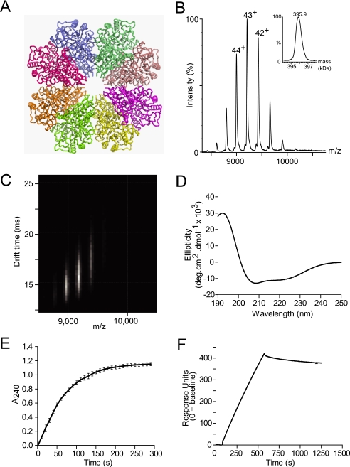
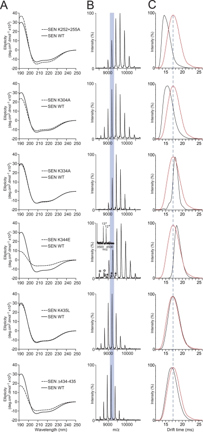
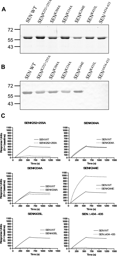
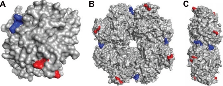
The flesh-eating bacterium group A Streptococcus (GAS) binds and activates human plasminogen, promoting invasive disease. Streptococcal surface enolase (SEN), a glycolytic pathway enzyme, is an identified plasminogen receptor of GAS. Here we used mass spectrometry (MS) to confirm that GAS SEN is octameric, thereby validating in silico modeling based on the crystal structure of Streptococcus pneumoniae alpha-enolase. Site-directed mutagenesis of surface-located lysine residues (SEN(K252 + 255A), SEN(K304A), SEN(K334A), SEN(K344E), SEN(K435L), and SEN(Delta434-435)) was used to examine their roles in maintaining structural integrity, enzymatic function, and plasminogen binding. Structural integrity of the GAS SEN octamer was retained for all mutants except SEN(K344E), as determined by circular dichroism spectroscopy and MS. However, ion mobility MS revealed distinct differences in the stability of several mutant octamers in comparison with wild type. Enzymatic analysis indicated that SEN(K344E) had lost alpha-enolase activity, which was also reduced in SEN(K334A) and SEN(Delta434-435). Surface plasmon resonance demonstrated that the capacity to bind human plasminogen was abolished in SEN(K252 + 255A), SEN(K435L), and SEN(Delta434-435). The lysine residues at positions 252, 255, 434, and 435 therefore play a concerted role in plasminogen acquisition. This study demonstrates the ability of combining in silico structural modeling with ion mobility-MS validation for undertaking functional studies on complex protein structures.


Figures

FIGURE 1.
FIGURE 1.
Structural and biochemical analysis, molecular modeling, and plasminogen binding ability of recombinant wild-type SEN. A, ribbon diagram of the model of wild-type SEN octamer modeled from the crystal structure of octameric S. pneumoniae α-enolase. B and C, ESI-MS (inset: spectrum transformed to mass scale) (B) and IM-MS plots showing the octameric structure of recombinant wild-type SEN (C). D, far-UV circular dichroism spectrum demonstrating the predominantly α-helical secondary structure of recombinant wild-type SEN, as shown by the minima at 209 and 222 nm. E, enzymatic activity of recombinant wild-type SEN. The graph shown was constructed from the averages of three independent experiments, with error bars indicating the standard deviation. deg, degree. F, representative surface plasmon resonance sensorgram for recombinant wild-type SEN (100 nm, as monomer) binding to human Glu-plasminogen; 10,200 response units of ligand had been immobilized on a CM5 chip using amine-coupling chemistry.
FIGURE 2.
FIGURE 2.
Structural analysis of recombinant SEN mutants. A, far-UV circular dichroism spectra comparing wild-type (WT) SEN (solid lines) and each of the mutant forms (dashed lines). deg, degree. B, ESI-mass spectra illustrating the major charge state series for each of the mutant octamers, with the 43+ charge state highlighted. SENK344E was observed to contain minor amounts of heptamer (*) and monomer (o) (shown in insets). C, IM-mass spectra showing the drift time for the 43+ charge state of each mutant (black profiles) when compared with that of wild-type SEN (red profiles).
FIGURE 3.
FIGURE 3.
Plasminogen binding ability of SEN mutants. A, Coomassie Brilliant Blue-stained 12% SDS-PAGE reducing gel of SEN wild type (WT) and mutants. B, corresponding ligand blot. Proteins were transferred to a membrane, incubated with Glu-plasminogen, and probed with antiserum specific for human plasminogen. C, surface plasmon resonance sensorgrams for recombinant wild-type and mutant SEN proteins binding at 100 nm (as monomer) to immobilized human Glu-plasminogen (10,200 response units).
FIGURE 4.
FIGURE 4.
Homology model of SEN based on the crystal structure of S. pneumoniae α-enolase showing lysine residues implicated in plasminogen binding. A, monomeric subunit. B, octameric structure. C, side view of octameric structure. Internal lysine residues at positions 252 and 255 are colored red, and C-terminal lysines are colored blue.

Similar articles

Cited by

References

    1. Carapetis J. R., Steer A. C., Mulholland E. K., Weber M. ( 2005) Lancet Infect. Dis. 5, 685– 694 - PubMed
    1. Cunningham M. W. ( 2000) Clin. Microbiol. Rev. 13, 470– 511 - PMC - PubMed
    1. Tart A. H., Walker M. J., Musser J. M. ( 2007) Trends Microbiol. 15, 318– 325 - PubMed
    1. Carapetis J. R., Walker A. M., Hibble M., Sriprakash K. S., Currie B. J. ( 1999) Epidemiol. Infect. 122, 59– 65 - PMC - PubMed
    1. McArthur J. D., McKay F. C., Ramachandran V., Shyam P., Cork A. J., Sanderson-Smith M. L., Cole J. N., Ringdahl U., Sjöbring U., Ranson M., Walker M. J. ( 2008) FASEB J. 22, 3146– 3153 - PubMed

Publication types

MeSH terms

LinkOut - more resources