Characterization of a unique class C acid phosphatase from Clostridium perfringens
- PMID: 19363079
- PMCID: PMC2687270
- DOI: 10.1128/AEM.01599-08
Characterization of a unique class C acid phosphatase from Clostridium perfringens
Abstract
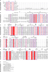
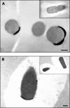
Clostridium perfringens is a gram-positive anaerobe and a pathogen of medical importance. The detection of acid phosphatase activity is a powerful diagnostic indicator of the presence of C. perfringens among anaerobic isolates; however, characterization of the enzyme has not previously been reported. Provided here are details of the characterization of a soluble recombinant form of this cell-associated enzyme. The denatured enzyme was approximately 31 kDa and a homodimer in solution. It catalyzed the hydrolysis of several substrates, including para-nitrophenyl phosphate, 4-methylumbelliferyl phosphate, and 3' and 5' nucleoside monophosphates at pH 6. Calculated K(m)s ranged from 0.2 to 0.6 mM with maximum velocity ranging from 0.8 to 1.6 micromol of P(i)/s/mg. Activity was enhanced in the presence of some divalent cations but diminished in the presence of others. Wild-type enzyme was detected in all clinical C. perfringens isolates tested and found to be cell associated. The described enzyme belongs to nonspecific acid phosphatase class C but is devoid of lipid modification commonly attributed to this class.
Figures


Similar articles
-
Genetic and biochemical analysis of a class C non-specific acid phosphatase (NSAP) of Clostridium perfringens.Microbiology (Reading). 2010 Jan;156(Pt 1):167-173. doi: 10.1099/mic.0.030395-0. Epub 2009 Oct 15. Microbiology (Reading). 2010. PMID: 19833778
-
Rapid confirmation of Clostridium perfringens by using chromogenic and fluorogenic substrates.Appl Environ Microbiol. 2001 Sep;67(9):4382-4. doi: 10.1128/AEM.67.9.4382-4384.2001. Appl Environ Microbiol. 2001. PMID: 11526053 Free PMC article.
-
The class C acid phosphatase of Helicobacter pylori is a 5' nucleotidase.Protein Expr Purif. 2004 Jan;33(1):48-56. doi: 10.1016/j.pep.2003.08.020. Protein Expr Purif. 2004. PMID: 14680961
-
An acid phosphatase from Manihot glaziovii as an alternative to alkaline Phosphatase for molecular cloning experiments.Biotechnol Lett. 2005 Dec;27(23-24):1865-8. doi: 10.1007/s10529-005-3894-z. Biotechnol Lett. 2005. PMID: 16328981
-
Protein tyrosine phosphatase: enzymatic assays.Methods. 2005 Jan;35(1):2-8. doi: 10.1016/j.ymeth.2004.07.002. Methods. 2005. PMID: 15588980 Review.
Cited by
-
Structural basis of the inhibition of class C acid phosphatases by adenosine 5'-phosphorothioate.FEBS J. 2011 Nov;278(22):4374-81. doi: 10.1111/j.1742-4658.2011.08360.x. Epub 2011 Oct 10. FEBS J. 2011. PMID: 21933344 Free PMC article.
-
Expression, purification and crystallization of an atypical class C acid phosphatase from Mycoplasma bovis.Acta Crystallogr Sect F Struct Biol Cryst Commun. 2011 Oct 1;67(Pt 10):1296-9. doi: 10.1107/S1744309111031551. Epub 2011 Sep 30. Acta Crystallogr Sect F Struct Biol Cryst Commun. 2011. PMID: 22102051 Free PMC article.
-
Phylogenetic distribution, biogeography and the effects of land management upon bacterial non-specific Acid phosphatase Gene diversity and abundance.Plant Soil. 2018;427(1):175-189. doi: 10.1007/s11104-017-3301-2. Epub 2017 Jun 12. Plant Soil. 2018. PMID: 30996484 Free PMC article.
-
Isolation and characterization of a recombinant class C acid phosphatase from Sphingobium sp. RSMS strain.Biotechnol Rep (Amst). 2022 Feb 8;33:e00709. doi: 10.1016/j.btre.2022.e00709. eCollection 2022 Mar. Biotechnol Rep (Amst). 2022. PMID: 35242619 Free PMC article.
-
Characterization of an extremophile bacterial acid phosphatase derived from metagenomics analysis.Microb Biotechnol. 2024 Apr;17(4):e14404. doi: 10.1111/1751-7915.14404. Microb Biotechnol. 2024. PMID: 38588312 Free PMC article.
References
-
- Bateman, A., and M. Bycroft. 2000. The structure of a LysM domain from E. coli membrane-bound lytic murein transglycosylase D (MltD). J. Mol. Biol. 299:1113-1119. - PubMed
-
- Calderone, V., C. Forleo, M. Benvenuti, M. C. Thaller, G. M. Rossolini, and S. Mangani. 2006. The first structure of a bacterial class B acid phosphatase reveals further structural heterogeneity among phosphatases of the haloacid dehalogenase fold. J. Mol. Biol. 365:708-721. - PubMed
-
- Chance, D. L., C. A. Jensen, T. J. Reilly, and T. P. Mawhinney. 2005. Analysis of bacterial colony biology using microwave-assisted processing and LR white resin. Microsc. Microanal. 11(Suppl. 2):968-969.
MeSH terms
Substances
LinkOut - more resources
Full Text Sources
Molecular Biology Databases

