. 2009 Jun;150(2):646-57.
doi: 10.1104/pp.109.138073. Epub 2009 Apr 10.

Mitochondrial and nuclear localization of a novel pea thioredoxin: identification of its mitochondrial target proteins

Affiliations

Mitochondrial and nuclear localization of a novel pea thioredoxin: identification of its mitochondrial target proteins

María C Martí et al. Plant Physiol. 2009 Jun.

Abstract

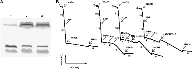
Plants contain several genes encoding thioredoxins (Trxs), small proteins involved in the regulation of the activity of many enzymes through dithiol-disulfide exchange. In addition to chloroplastic and cytoplasmic Trx systems, plant mitochondria contain a reduced nicotinamide adenine dinucleotide phosphate-dependent Trx reductase and a specific Trx o, and to date, there have been no reports of a gene encoding a plant nuclear Trx. We report here the presence in pea (Pisum sativum) mitochondria and nuclei of a Trx isoform (PsTrxo1) that seems to belong to the Trx o group, although it differs from this Trx type by its absence of introns in the genomic sequence. Western-blot analysis with isolated mitochondria and nuclei, immunogold labeling, and green fluorescent protein fusion constructs all indicated that PsTrxo1 is present in both cell compartments. Moreover, the identification by tandem mass spectrometry of the native mitochondrial Trx after gel filtration using the fast-protein liquid chromatography system of highly purified mitochondria and the in vitro uptake assay into isolated mitochondria also corroborated a mitochondrial location for this protein. The recombinant PsTrxo1 protein has been shown to be reduced more effectively by the Saccharomyces cerevisiae mitochondrial Trx reductase Trr2 than by the wheat (Triticum aestivum) cytoplasmic reduced nicotinamide adenine dinucleotide phosphate-dependent Trx reductase. PsTrxo1 was able to activate alternative oxidase, and it was shown to interact with a number of mitochondrial proteins, including peroxiredoxin and enzymes mainly involved in the photorespiratory process.


Figures

Figure 1.
Figure 1.
Amino acid sequence analysis of PsTrxo1. A, Sequence alignment of pea PsTrxo1, Arabidopsis AtTrxo1, and poplar PtTrxh2 using the ClustalW software. Catalytic sites are underlined. The transit peptide predicted by the MitoProt program in PsTrxo1 is shown in italics, and the anti-PsTrxo1 antibody produced against the peptide is shown in boldface. B, Phylogenetic tree of Arabidopsis Trxs and Trx o homologs from selected plant species (TreeView program). Protein sequences were obtained from the database or deduced from cDNA or EST sequences. The three described mitochondrial Trxs are circled.
Figure 2.
Figure 2.
Activity of recombinant PsTrxo1 determined by the insulin-disulfide reduction assay. The reduction of insulin was measured by the increase in turbidity at 650 nm for 1 h at 30°C. Triangles, wheat NADPH/TR; circles, S. cerevisiae Trr2.
Figure 3.
Figure 3.
Subcellular location of PsTrxo1 in pea leaves. A, Western-blot analysis of recombinant PsTrxo1 (r), purified mitochondria (Mit.), nuclei (Nucl.), chloroplasts (Chl.), and cytoplasm (Cyt.) from pea leaves using the antibody generated against the peptide of the PsTrxo1 amino acid sequence indicated in Figure 1A. B, Immunolocalization of PsTrxo1 in pea leaves by transmission electronic microscopy. Left, Nonstained nucleus with detail below; right, mitochondria stained with lead citrate and uranyl acetate. The table shows the gold particles found in each cellular compartment. Eu., Euchromatin; Het., heterochomatin.
Figure 4.
Figure 4.
Targeting of PsTrxo1 protein. A, Import of precursor PsTrxo1-35S into pea chloroplasts and potato mitochondria. Soybean AOX and pea small subunit of Rubisco (SSU) were used as controls. Lanes 1 and 4, Precursor protein alone; lane 2, precursor protein incubated with isolated chloroplasts; lane 3, as for lane 2 but with the addition of thermolysin after the import; lane 5, precursor protein incubated with isolated mitochondria; lane 6, as for lane 5 but with the addition of proteinase K after the import; lane 7, as for lane 5 but with the addition of valinomycin before the import; lane 8, as for lane 7 but with the addition of proteinase K after the import. Migration of the precursor (p) and mature (m) polypeptides is indicated. B, Subcellular location of PsTrxo1 protein in onion epidermal cells visualized by confocal microscopy. Epidermal onion layers were transiently transformed with the PsTrxo1 gene fused to GFP (panels 1–8) under the control of the 35S cauliflower mosaic virus promoter. After incubation for 24 h, cells were observed with a confocal microscope for emission spectrum of the GFP (panels 1 and 5). As a control for location, MitoTracker (panel 2) and DAPI staining (panel 6) as well as Nomarski observations (panels 4 and 8) were carried out. Panel 3 depicts the merged images from panels 1 and 2, and panel 7 depicts the merged images from panels 6 and 8. Arrows in panels 1 to 3 indicate mitochondria, and arrows in panels 5 to 7 indicate nuclei. As a control, the transformed cells with the reporter gene GFP (panels 9 and 10) are shown.
Figure 5.
Figure 5.
Identification of the native mitochondrial PsTrxo1. A, Superdex 200 filtration chromatography of purified mitochondrial proteins. B and C, SDS-PAGE (B) and western-blot analysis (C) of recombinant PsTrxo1 (lane 1) and concentrated low molecular mass fractions indicated between arrows in A (lane 2). Molecular mass markers are shown in lane 3.
Figure 6.
Figure 6.
Regulation of AOX by PsTrxo1. A, AOX reduction. Lane 1, Pea mitochondria were incubated with the NADPH/Trr2/PsTrxo1 system and subjected to SDS-PAGE; lane 2, as for lane 1 but without the addition of PsTrxo1; lane 3, control mitochondria. AOX dimers and monomers were immunodetected. B, AOX activation. Oxygen uptake by pea mitochondria previously treated with 10 mm diamide was measured in a Suc-depleted medium. Trace 1, NADH (2 mm) oxidation by purified mitochondria; traces 2 to 4, where indicated, different components were added to the oxygen electrode chamber. The Trx system was NADPH/Trr2/PsTrxo1 (trace 3). As controls, 5 mm DTT (trace 2) or NADPH/Trr2 (trace 4) was added. Numbers on the traces represent nmol O2 min−1 mg−1 protein from one of three experiments. Mixot., Mixothiazol (inhibitor of cytochrome c oxidase); Pyr., sodium pyruvate; SHAM, salicylhydroxamic acid (inhibitor of AOX).

Similar articles

Cited by

References

    1. Alkhalfioui F, Renard M, Frendo P, Keichinger C, Meyer Y, Gelhaye E, Hirasawa M, Knaff DB, Ritzenthaler C, Montrichard F (2008) A novel type of thioredoxin dedicated to symbiosis in legumes. Plant Physiol 148 424–435 - PMC - PubMed
    1. Altschul SF, Madden TL, Schaffer AA, Zhang J, Zhang Z, Miller W, Lipman DJ (1997) Gapped BLAST and PSI-BLAST: a new generation of protein database search programs. Nucleic Acids Res 25 3389–3402 - PMC - PubMed
    1. Anderson S, Smith SM (1986) Synthesis of the small subunit of ribulose-bisphosphate carboxylase from genes cloned into plasmids containing the sp6 promoter. Biochem J 240 709–715 - PMC - PubMed
    1. Arai RJ, Masutani H, Yodoi J, Debbas V, Laurindo FR, Stern A, Monteiro HP (2006) Nitric oxide induces thioredoxin-1 nuclear translocation: possible association with the p21Ras survival pathway. Biochem Biophys Res Commun 348 1254–1260 - PubMed
    1. Arner ESJ, Holmgren A (2000) Physiological functions of thioredoxin and thioredoxin reductase. Eur J Biochem 267 6102–6109 - PubMed

Publication types

MeSH terms

Associated data

LinkOut - more resources