. 2009 Apr 21;106(16):6650-5.
doi: 10.1073/pnas.0901083106. Epub 2009 Apr 10.

Beta-arrestin-dependent signaling and trafficking of 7-transmembrane receptors is reciprocally regulated by the deubiquitinase USP33 and the E3 ligase Mdm2

Affiliations

Beta-arrestin-dependent signaling and trafficking of 7-transmembrane receptors is reciprocally regulated by the deubiquitinase USP33 and the E3 ligase Mdm2

Sudha K Shenoy et al. Proc Natl Acad Sci U S A. .

Abstract

Beta-arrestins are multifunctional adaptors that mediate the desensitization, internalization, and some signaling functions of seven-transmembrane receptors (7TMRs). Agonist-stimulated ubiquitination of beta-arrestin2 mediated by the E3 ubiquitin ligase Mdm2 is critical for rapid beta(2)-adrenergic receptor (beta(2)AR) internalization. We now report the discovery that the deubiquitinating enzyme ubiquitin-specific protease 33 (USP33) binds beta-arrestin2 and leads to the deubiquitination of beta-arrestins. USP33 and Mdm2 function reciprocally and favor respectively the stability or lability of the receptor beta-arrestin complex, thus regulating the longevity and subcellular localization of receptor signalosomes. Receptors such as the beta(2)AR, previously shown to form loose complexes with beta-arrestin ("class A") promote a beta-arrestin conformation conducive for binding to the deubiquitinase, whereas the vasopressin V2R, which forms tight beta-arrestin complexes ("class B"), promotes a distinct beta-arrestin conformation that favors dissociation of the enzyme. Thus, USP33-beta-arrestin interaction is a key regulatory step in 7TMR trafficking and signal transmission from the activated receptors to downstream effectors.


Conflict of interest statement

The authors declare no conflict of interest.

Figures

Fig. 1.
Fig. 1.
Identification of USP33 as a β-arrestin-binding deubiquitinase. (A) HA-USP33 transiently expressed in COS-7 cells along with β-arrestin1-Flag, β-arrestin2-Flag, or vector was immunoprecipitated (IP) with anti-hemagglutinin epitope (HA)-agarose conjugate, and the amount of bound β-arrestin1 and -2 was assessed by probing with an anti-β-arrestin antibody (Upper). IB, immunoblot. Expression levels of USP33 and β-arrestins in cell extracts are also displayed (Lower). Data shown are from 1 of 4 independent experiments. (B) The bands displayed represent purified USP33 that is either unmodified or covalently linked to ubiquitin-vinyl sulfone (Ub-VS), separated on an SDS/polyacrylamide gel (6%), and detected by Sypro ruby red staining. (C) Lys-63-linked polyubiquitin chains, which lack monoubiquitin, were incubated with buffer (lane 1), Isopeptidase T (lane 2), or increasing amounts of purified USP33 (lanes 3–5). The appearance of a monoubiquitin band corresponds to the depolymerizing activity of USPs. Data displayed in B and C are representative of 3 or 4 experiments performed with 3 separate USP33 purifications.
Fig. 2.
Fig. 2.
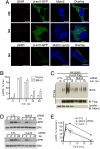
USP33 inhibits vasopressin-stimulated β-arrestin ubiquitination, endosomal trafficking, and ERK activation. (A) Flag immunoprecipitates isolated before or after 1 μM [Arg]vasopressin (AVP) stimulation from HEK-293 cells that transiently express the V2R with either pcDNA3 or β-arrestin2-Flag were treated with a mock purification (first 4 lanes in each gel) or purified HA-USP33 (last 4 lanes in each gel). After incubation, samples were subjected to Western analyses with an anti-ubiquitin antibody (Left) and reprobed with an anti-Flag antibody (Right). (B) HEK-293 cells with stable HA-V2R expression were transiently transfected with β-arrestin2-GFP and RFP-USP33 and stimulated with 1 μM AVP for 20 min. Two cells are shown, one with no detectable RFP-USP33 that has the expected endosomal recruitment of β-arrestin2, the other that expresses USP33 in which β-arrestin recruitment to the endosomes is inhibited. (C) Confocal micrographs show endosomal distribution of β-arrestin2-GFP upon AVP stimulation in cells overexpressing USP33Cys:His. (D) HEK-293 cells transiently expressing HA-V2R with either vector or USP33 were stimulated with 1 μM AVP for the indicated times and cell lysates were analyzed for ERK and phosphorylated ERK (p-ERK) by immunoblotting. (E) The graph represents quantification of p-ERK stimulated by a time course of AVP in HEK-293 transiently transfected with HA-V2R along with vector or USP33 from 4 independent experiments. *, P < 0.01 by 2-way ANOVA, n = 4.
Fig. 3.
Fig. 3.
Mdm2 promotes β-arrestin recruitment to endosomes and enhances signaling. (A) HEK-293 cells transiently expressing HA-β2AR and Mdm2 without stimulation (NS, Top) or with 20-min isoproterenol (Iso, Middle) stimulation are fixed and immunostained to detect the receptor (anti-β2AR, H-20) shown in red and Mdm2 (Ab-1) displayed in blue. Distribution of β-arrestin2-GFP is shown in green. In Bottom, confocal pictures of agonist-stimulated HEK-293 cells expressing HA-β2AR, but with endogenous (end.) Mdm2 are shown. (Scale bars, 10 μm.) (B) Bar graph showing quantification (n = 3) of p-ERK in whole-cell extracts in HEK-293 cells with or without exogenous Mdm2 expression in response to isoproterenol stimulation for indicated times. *, P < 0.05, 2-way ANOVA. (C) (Top) HEK-293 cells were transiently transfected with HA-β2AR and β-arrestin2-Flag along with siRNA that targets nothing (control, CTL) or Mdm2; 48–60 hr posttransfection, β-arrestin2 was immunoprecipitated after agonist treatment for the indicated times and the immunoprecipitate was probed with an anti-ubiquitin antibody. (Middle) The amount of β-arrestin in the immunoprecipitate. (Lower) The levels of Mdm2 in cell extracts. (D) HEK-293 cells with or without either Mdm2 or β-arrestin2 depletion are analyzed for isoproterenol-stimulated p-ERK and ERK. (E) Graph represents quantification of time course of p-ERK from 4 independent experiments ± SEM. Two-way ANOVA, **, P < 0.01; *, P < 0.05, CTL versus Mdm2, and CTL versus β-arr2, 5- and 20-min signals, respectively.
Fig. 4.
Fig. 4.
USP33 knockdown prevents β-arrestin deubiquitination, promotes endosomal trafficking, and prolongs ERK signaling. (A) HEK-293 cells were transfected with HA-β2AR, β-arrestin2-Flag, and either control or USP33 siRNA. Flag immunoprecipitates were isolated after isoproterenol stimulation for the indicated times and probed with an anti-ubiquitin antibody (Upper). (Lower) USP33 levels in control and USP-knockdown samples. (B) Confocal micrographs show distribution of Flag-β2AR (red) and β-arrestin2-GFP (green) in USP33-depleted cells (Upper) and control (Lower) cells in agonist-stimulated HEK-293 cells. (Scale bars, 10 μm.) (C) Western blots of p-ERK and ERK in response to a time course of isoproterenol (100 nM) activation, detected in HEK-293 cells with stable β2AR expression (2 pmol/mg of protein) and with indicated siRNA transfections. (D) Quantification of p-ERK signals from 3 independent experiments. **, P < 0.01, control versus USP33, 2-way ANOVA. (E) A representative blot for USP33 levels showing Western analysis of 25 μg of lysate protein from each siRNA transfection.
Fig. 5.
Fig. 5.
β-Arrestin–USP33 interaction displays different kinetics upon stimulation of different 7TMRs and is dependent on distinct conformational changes. (A) COS-7 cells transiently expressing either HA-β2AR or HA-V2R along with β-arrestin2-Flag were stimulated with respective agonists for the indicated times, β-arrestins were immunoprecipitated, and the immunoprecipitate was probed with USP33 antisera. The graphs show the quantification of USP33 bound to isolated β-arrestin obtained from 4 independent experiments. The 4 time points within each binding curve are analyzed by 1-way ANOVA. In each case, stimulated samples are significantly different from unstimulated samples; *, P < 0.05; **, P < 0.01; ***, P < 0.001. (B) Purified HA-USP33 was incubated alone or with β-arrestin2-His6 without or with indicated receptor peptides (see Methods). USP33 binding from 4 separate experiments is quantified and normalized to β-arrestin levels and plotted as bar graphs; #, P < 0.05, β2AR-PP versus β2AR-NP; **, P < 0.01, β2AR-PP versus all other samples, 1-way ANOVA, Bonferoni comparison. (C) SDS/PAGE analyses of the limited tryptic proteolytic products of β-arrestin2 with indicated receptor peptides (see Methods). Red arrows indicate the significant differences in the limited proteolysis patterns.
Fig. 6.
Fig. 6.
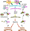
Schematic showing the effects of posttranslational modifications in 7TMR signaling. Step 1, β-arrestin2 resides in a basal state in the cytoplasm and is recruited to the plasma membrane and binds phosphorylated C termini of 7TMRs. The sites of phosphorylation differ among the 2 representative receptors shown. Step 2, upon binding to each receptor, β-arrestin2 undergoes a distinct conformational reorientation, thus allowing distinct regions to become modified by ubiquitination. Step 3, the β2AR-induced conformation promotes β-arrestin2–USP33 interaction. Step 4, USP33 deubiquitinates β-arrestin, leading to the dissociation of β-arrestin from the β2AR. Step 5, β2AR–β-arrestin2 signalosomes are short-lived and promote transient ERK activity that is predominantly nonendosomal. Step 6, β-arrestin2 conformation induced by the V2R activation prevents USP33 binding, thus protecting β-arrestin ubiquitination, allowing tight binding to activated receptors. Step 7, V2R–β-arrestin2 signalosomes are stable and result in robust ERK activity that is predominantly localized on endosomes.

References

    1. Drake MT, Shenoy SK, Lefkowitz RJ. Trafficking of G protein-coupled receptors. Circ Res. 2006;99:570–582. - PubMed
    1. DeWire SM, Ahn S, Lefkowitz RJ, Shenoy SK. Beta-arrestins and cell signaling. Annu Rev Physiol. 2007;69:483–510. - PubMed
    1. Oakley RH, et al. Differential affinities of visual arrestin, beta arrestin1, and beta arrestin2 for G protein-coupled receptors delineate two major classes of receptors. J Biol Chem. 2000;275:17201–17210. - PubMed
    1. Shenoy SK, McDonald PH, Kohout TA, Lefkowitz RJ. Regulation of receptor fate by ubiquitination of activated beta 2-adrenergic receptor and beta-arrestin. Science. 2001;294:1307–1313. - PubMed
    1. Shenoy SK, Lefkowitz RJ. Trafficking patterns of beta-arrestin and G protein-coupled receptors determined by the kinetics of beta-arrestin deubiquitination. J Biol Chem. 2003;278:14498–14506. - PubMed

Publication types

MeSH terms