. 2009 May;16(5):528-33.
doi: 10.1038/nsmb.1577. Epub 2009 Apr 12.

Insights into substrate stabilization from snapshots of the peptidyl transferase center of the intact 70S ribosome

Affiliations

Insights into substrate stabilization from snapshots of the peptidyl transferase center of the intact 70S ribosome

Rebecca M Voorhees et al. Nat Struct Mol Biol. 2009 May.

Abstract

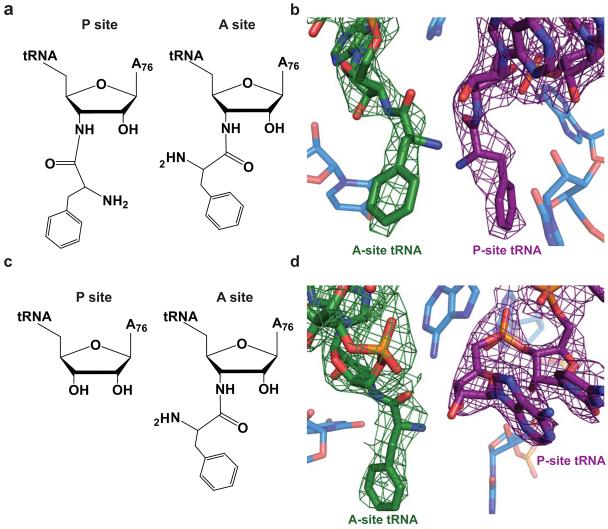
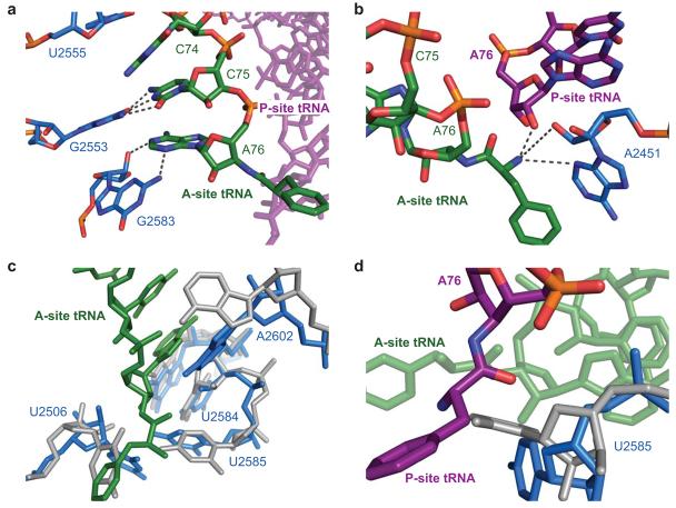
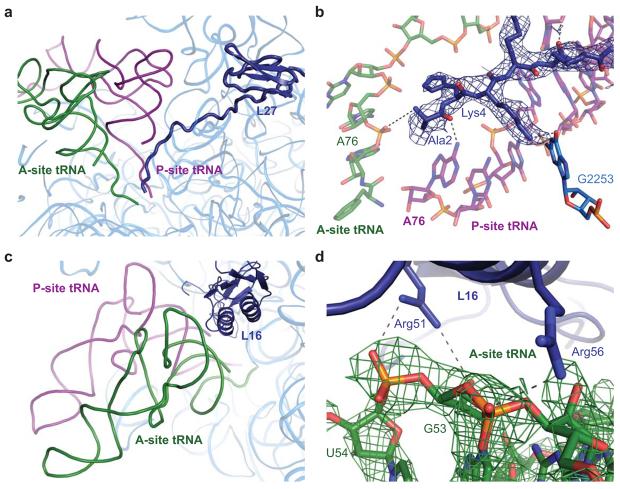
Protein synthesis is catalyzed in the peptidyl transferase center (PTC), located in the large (50S) subunit of the ribosome. No high-resolution structure of the intact ribosome has contained a complete active site including both A- and P-site tRNAs. In addition, although past structures of the 50S subunit have found no ordered proteins at the PTC, biochemical evidence suggests that specific proteins are capable of interacting with the 3' ends of tRNA ligands. Here we present structures, at 3.6-A and 3.5-A resolution respectively, of the 70S ribosome in complex with A- and P-site tRNAs that mimic pre- and post-peptidyl-transfer states. These structures demonstrate that the PTC is very similar between the 50S subunit and the intact ribosome. They also reveal interactions between the ribosomal proteins L16 and L27 and the tRNA substrates, helping to elucidate the role of these proteins in peptidyl transfer.


Figures

Figure 1
Figure 1
Ribosomal substrates in the peptidyl transferase center. a) Chemical diagram of the pre-peptidyl transfer state of the ribosomal active site. In this structure both the A- and P-site tRNAs contain an amide linkage between residue A76 and the phenylalanine amino acid. b) Model of the ribosomal active site in the pre-peptidyl transfer state including representative 3Fo-2Fc density for the A- and P-site tRNAs in green and purple, respectively. c) Chemical diagram of the post-peptidyl transfer state in which the A site contains an amide-linked Phe-tRNAPhe and the P site contains tRNAfMet. d) Model of the post-peptidyl transfer in the peptidyl transferase center including 3Fo-2Fc density for the A- and P-site tRNAs in green and purple, respectively.
Figure 2
Figure 2
Interactions of the ribosomal substrates with the 23S RNA in the peptidyl transferase center. a) Model of the ribosome and the stabilizing interactions between the CCA tail of A-site tRNA, shown in green, and the 23S RNA, shown in light blue as determined in the pre-peptidyl transfer structure. b) Interactions of the A-site amino acid backbone with the 23S RNA, shown in blue, and the peptidyl-tRNA, shown in purple. For ease of illustration, the structure of the post-peptidyl transfer state of the ribosome is displayed. c) Superposition of the 23S RNA from Selmer et al. (displayed in gray), which contains an empty 50S A site, with that of the pre-peptidyl transfer structure presented here, containing an occupied A site (displayed in blue). Shifts in residues U2584, U2585, U2506, and A2602 are characteristic of A-site accommodation. d) Model of the conformational change of residue U2585 that, upon binding of A-site tRNA, exposes the peptidyl-tRNA ester for nucleophilic attack.
Figure 3
Figure 3
Interactions of the ribosomal proteins L27 and L16 with the ribosomal substrates. a) Overview of protein L27 in relation to the A- and P-site tRNAs, shown in green and purple, respectively. The protein, shown in dark blue, contains a globular domain and an N-terminal extension that localizes between the 3′ ends of the ribosomal tRNAs. b) Predicted interactions of protein L27 with the ribosomal substrates and 23S RNA (shown in light blue). The modeled interactions were observed in both structures containing occupied A sites, though the post-peptidyl transfer structure is displayed here as it contained moderately better electron density for L27. A representative 3Fo-2Fc electron density map is displayed in blue. c) Overview of protein L16 in relation to the ribosomal substrates. The protein is located adjacent to the elbow of the A-site tRNA. d) Interactions between the conserved residues Arg51 and Arg56 of protein L16, shown in dark blue, with the backbone of the A-site tRNA, shown in green. Representative 3Fo-2Fc density, as determined in the pre-peptidyl transfer structure, is displayed in green for the region of the A-site tRNA predicted to interact with L16.

Similar articles

Cited by

References

    1. Nissen P, Hansen J, Ban N, Moore PB, Steitz TA. The structural basis of ribosome activity in peptide bond synthesis. Science. 2000;289:920–30. - PubMed
    1. Steitz TA. Structural insights into the functions of the large ribosomal subunit, a major antibiotic target. Keio J Med. 2008;57:1–14. - PubMed
    1. Rodnina MV, Beringer M, Wintermeyer W. Mechanism of peptide bond formation on the ribosome. Q Rev Biophys. 2006;39:203–25. - PubMed
    1. Korostelev A, Trakhanov S, Laurberg M, Noller HF. Crystal structure of a 70S ribosome-tRNA complex reveals functional interactions and rearrangements. Cell. 2006;126:1065–77. - PubMed
    1. Bashan A, et al. Structural basis of the ribosomal machinery for peptide bond formation, translocation, and nascent chain progression. Mol Cell. 2003;11:91–102. - PubMed

Publication types