. 2009 May;11(5):604-15.
doi: 10.1038/ncb1866. Epub 2009 Apr 12.

Persistent transcription-blocking DNA lesions trigger somatic growth attenuation associated with longevity

Affiliations

Persistent transcription-blocking DNA lesions trigger somatic growth attenuation associated with longevity

George A Garinis et al. Nat Cell Biol. 2009 May.

Abstract

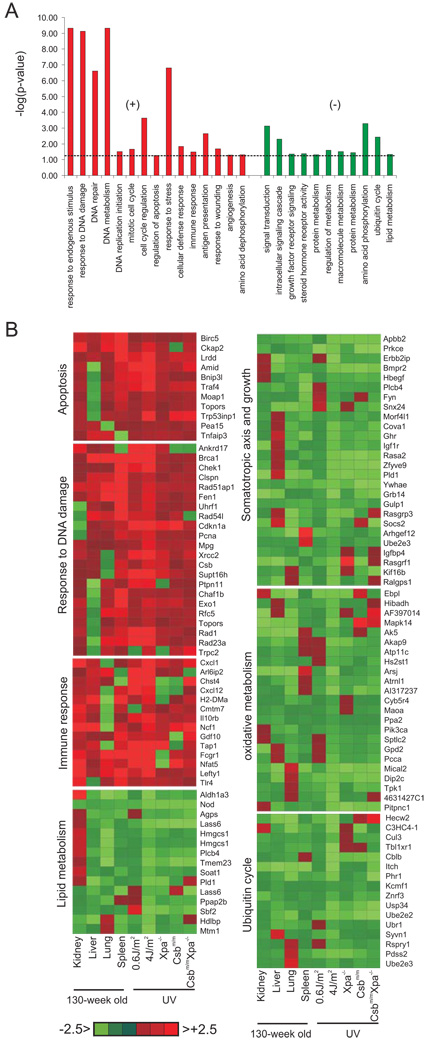
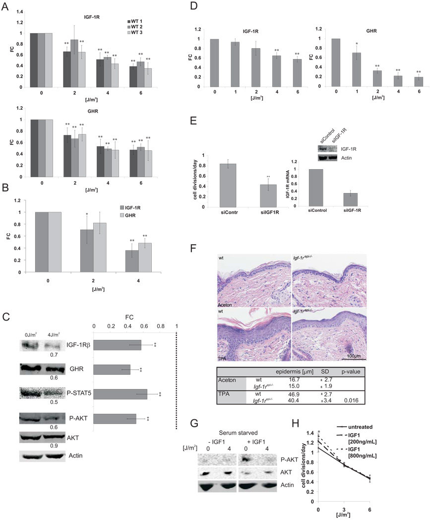
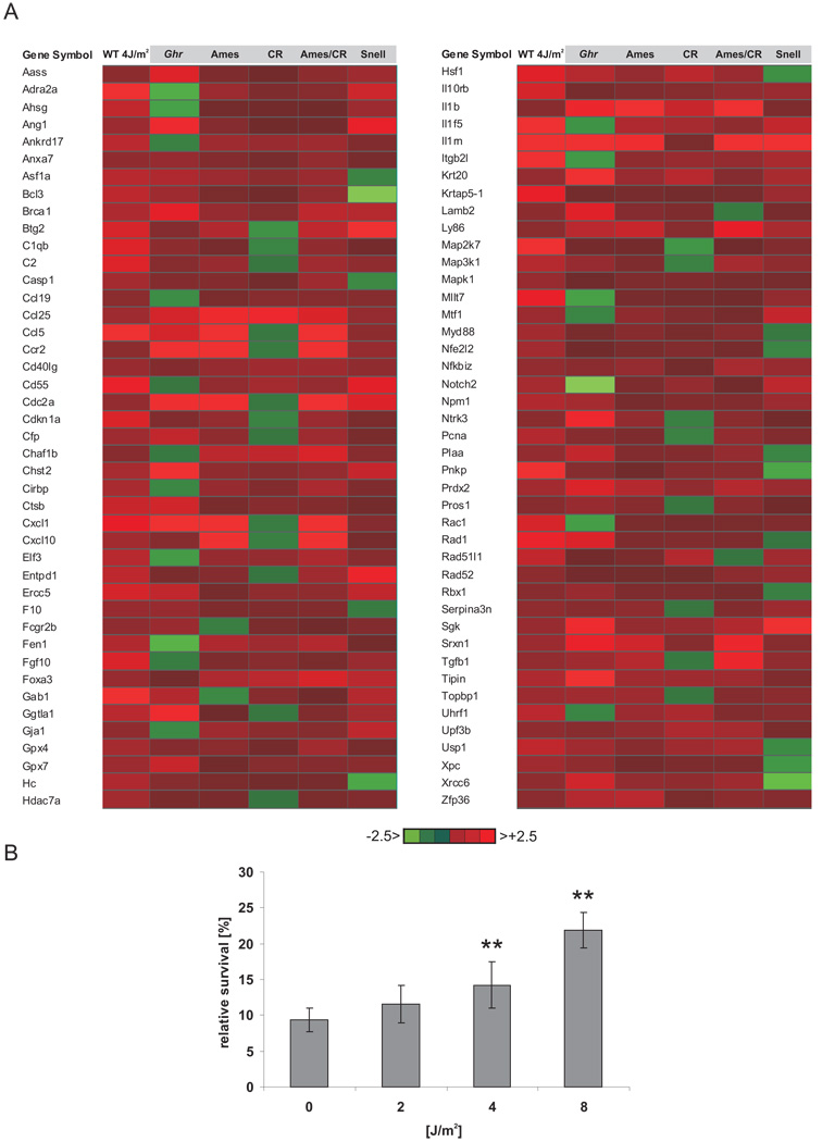
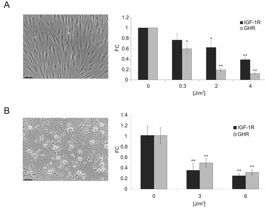
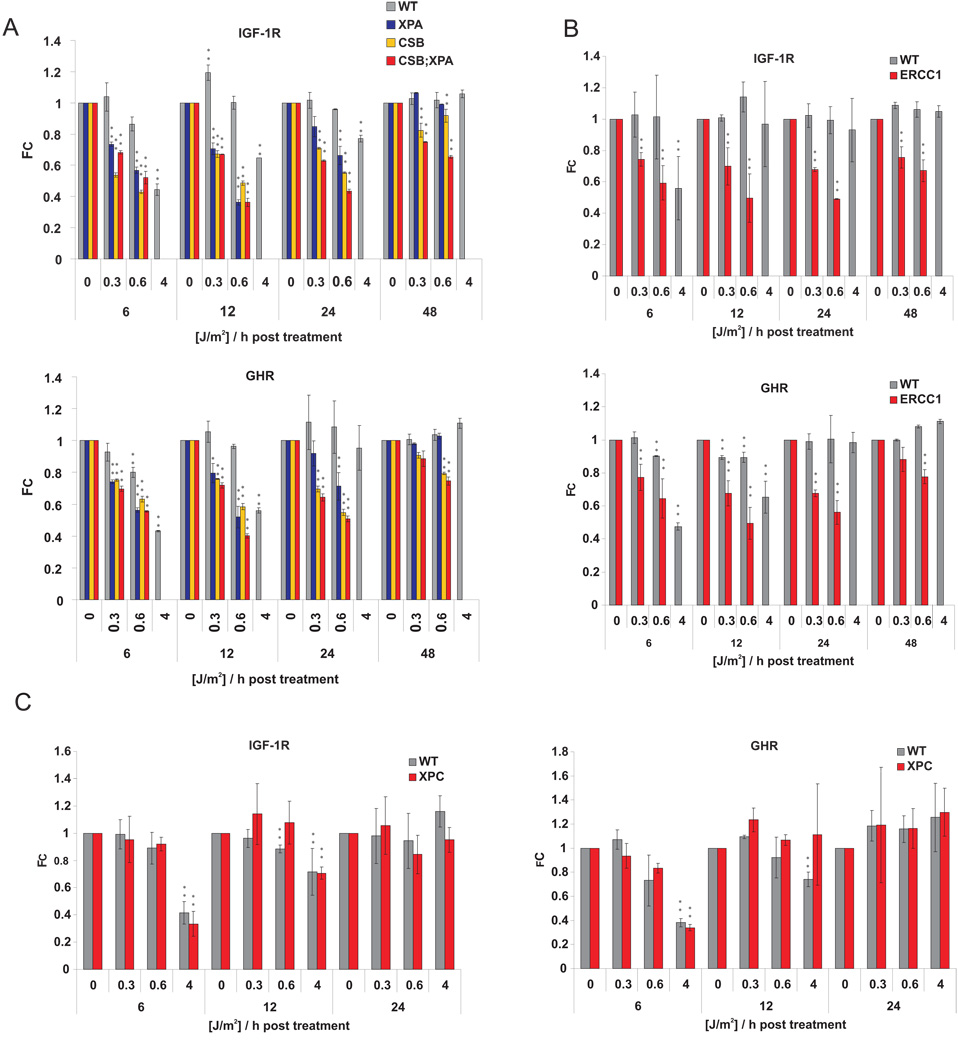
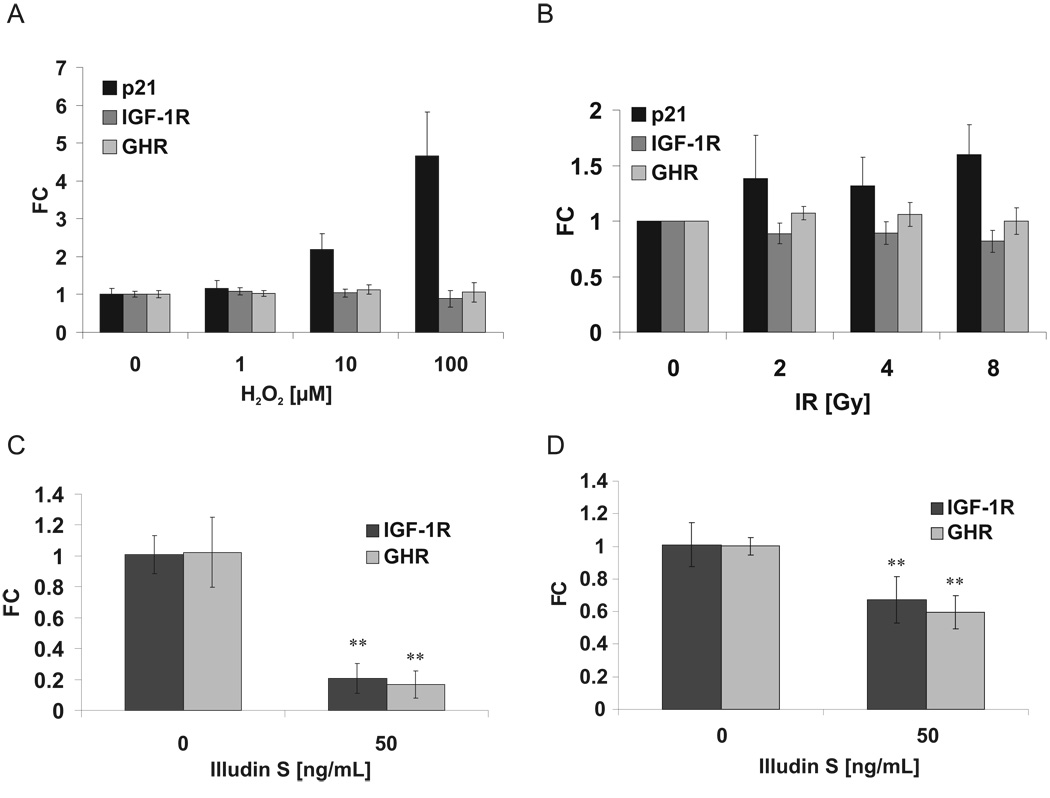
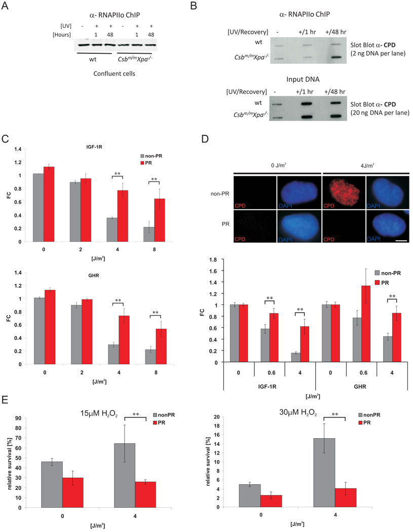
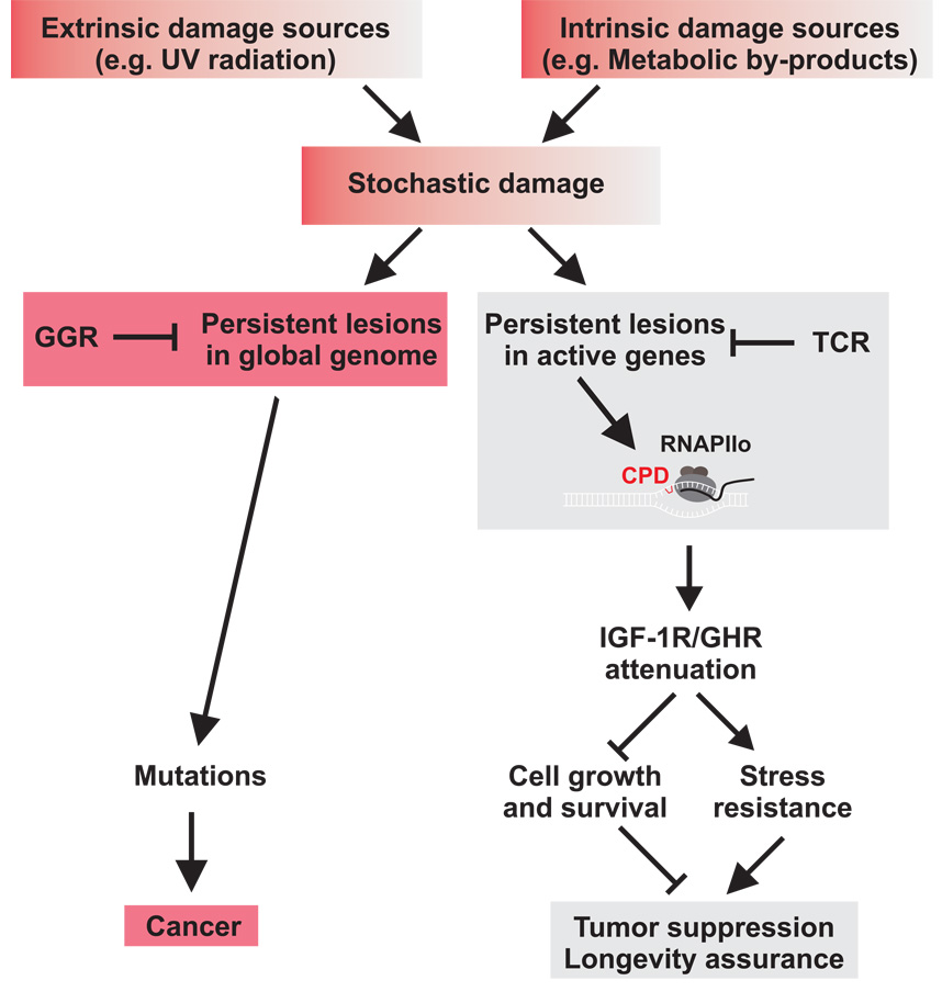
The accumulation of stochastic DNA damage throughout an organism's lifespan is thought to contribute to ageing. Conversely, ageing seems to be phenotypically reproducible and regulated through genetic pathways such as the insulin-like growth factor-1 (IGF-1) and growth hormone (GH) receptors, which are central mediators of the somatic growth axis. Here we report that persistent DNA damage in primary cells from mice elicits changes in global gene expression similar to those occurring in various organs of naturally aged animals. We show that, as in ageing animals, the expression of IGF-1 receptor and GH receptor is attenuated, resulting in cellular resistance to IGF-1. This cell-autonomous attenuation is specifically induced by persistent lesions leading to stalling of RNA polymerase II in proliferating, quiescent and terminally differentiated cells; it is exacerbated and prolonged in cells from progeroid mice and confers resistance to oxidative stress. Our findings suggest that the accumulation of DNA damage in transcribed genes in most if not all tissues contributes to the ageing-associated shift from growth to somatic maintenance that triggers stress resistance and is thought to promote longevity.


Figures

Figure 1
Figure 1. Shared biological processes upon induction of DNA damage in vitro and aging in vivo
A) List of biological processes that are significantly over-represented upon induction of DNA damage and during aging (left panel: upregulated; right panel: downregulated). B) Expression profiles of genes associated with the significantly over-represented biological processes upon UV treatment in vitro and in aging tissues. For each gene, the average expression value of UV-exposed primary MDFs (4 independent MDF lines were used for each genotype (n=4)) and the indicated tissue from 130-week old mice is compared to that of the corresponding non-treated cells or tissue of 13-week old young mice (average of 3 old vs. 3 young mice (n=3)), respectively. Deeper red or green colors indicate positive and negative fold changes respectively.
Figure 2
Figure 2. IGF-1R and GHR are repressed upon persistent DNA damage and leads to IGF-1 resistance
A) Three independent primary mouse dermal fibroblast cell lines (WT1–3; n=3)) at early passage were treated with UV (UV-C, 254 nm) and RNA samples were taken 6 hrs post treatment. IGF-1R and GHR qPCRs were done in duplicate and normalized to gamma-tubulin (gTUB) and beta-2-microglobulin (B2M). B) Primary chondrocytes were treated similarly and IGF-1R and GHR expression levels normalized to B2M, gTUB, and Hprt (average of two independent chondrocyte lines shown) and (C) IGF-1R and GHR protein and phosphorylation levels of STAT5 and AKT were quantified 12 h after UV treatment using fluorescently labeled antibodies and normalized to Actin levels (left panels: representative experiment; right panel: average of three independent experiments, n=3). D) Two independent human fibroblast lines (C5RO and C3RO) were treated with indicated doses of UV. IGF-1R and GHR expression levels were determined 6 hrs post treatment and normalized to Gapdh and B2M. E) IGF-1R down-regulation is sufficient to reduce cell growth. U2OS cells were counted 96 hours after transfection with either non-targeting siRNA oligos or siRNA oligos targeting IGF-1R (daily division rate given, n=4; upper right panel IGF-1R protein levels, lower right panel IGF-1R mRNA levels (qPCR)). F) IGF-1R ablation antagonizes hyperplasia. 8 – 10 weeks old Igf-1repi−/− and control mice were shaved and after 24 hours, treated three times every 48 hours with 10nm TPA/330µl or solvent alone (aceton). Upper panel: H&E section showing epidermal hyperplasia following TPA treatments. Lower panel: Quantification of hyperproliferative response (TPA-treated: n = 3–5, aceton treated: n = 2; two-tailed t-test). UV induces IGF-1 resistance: (G) U2OS cells were serum starved for 2 hours starting at 12 hours after UV treatment and treated with 100ng/mL IGF1 for 10 minutes. H) Medium was supplemented with IGF-1 after UV treatment and cells were counted 48 hours after treatment. (FC = fold change compared to untreated controls; *=p<0.05, **=p<0.01, two-tailed t-test, error bars = SD, n=4)
Figure 3
Figure 3. UV response induces oxidative stress resistance
A) Common stress responses are induced in response to UV damage in primary cells and in pituitary dwarf, Ghr knockout and calorie restricted mice with extended longevity. B) U2OS cells were treated with indicated UV doses and 12h post radiation treated with 100µM hydrogen peroxide. Relative survival compared to non-hydrogen peroxide treated cells (*=p<0.05, **=p<0.01, two-tailed t-test, error bars = SD, n=4).
Figure 4
Figure 4. UV irradiation leads to IGF-1R and GHR attenuation in quiescent and terminally differentiated cells
A) Quiescent C5RO primary human fibroblasts were treated 6 days after transfer to 1% FCS medium and samples taken 6hrs post UV treatment. IGF-1R and GHR expression levels were normalized to Gapdh and gTUB. B) Primary rat neurons were treated with UV and IGF-1R and GHR expression was normalized to Hprt and Gapdh (FC = fold change compared to untreated controls; *=p<0.05, **=p<0.01, two-tailed t-test, error bars = SD; n=4; scale bar 25µm).
Figure 5
Figure 5. Prolonged IGF-1R and GHR repression in cells from NER deficient progeroid mice
(A), Csbm/m/Xpa−/−, Csbm/m and Xpa−/−, (B) Ercc1−/−, (C) Xpc−/− and wt littermate control primary MDFs at early passage were treated with indicated UV doses and samples taken at indicated time points after treatment. IGF-1R and GHR expression levels were evaluated by qPCR and normalized to gTUB and B2M. (FC = fold change compared to untreated controls; *=p<0.05, **=p<0.01, two-tailed t-test, error bars = SD, n=4).
Figure 6
Figure 6. Attenuation of IGF1-R and GHR expression in response to illudin S but not to oxidative damage and ionizing irradiation
Primary MDFs were treated with hydrogen peroxide (A) or ionizing radiation (B) and IGF-1R and GHR expression levels were evaluated after 6 h by qPCR and normalized to gTUB and B2M. p21 expression was included as control for DNA damage response. Illudin S treatment leads to IGF-1R and GHR attenuation in quiescent human fibroblasts (C) and rat neurons (D), normalized to Gapdh and gTUB (C) or Hprt and Gapdh (D) (FC = fold change compared to untreated controls; **=p<0.01, two-tailed t-test, error bars = SD; n=4).
Figure 7
Figure 7. Repair of persistent CPD lesions alleviates IGF-1R and GHR repression and oxidative stress resistance
A) RNAPIIo protein levels in RNAPIIo specific ChIP using chromatin of crosslinked UV- and non-irradiated confluent chondrocytes (as indicated). B) Slot blot analysis of different amounts of RNAPIIo co-ChIPed DNA (top panel) and input crosslinked DNA (lower panel) spotted at the same membrane using a CPD specific antibody (representative experiment shown). CPD and 6-4PP photolyase transgenic MEFs (C) or a CPD photolyase transgenic CS1AN human CS cell line (D) were UV treated and then either photoreactivated (PR) for 1h or kept in the dark (non-PR). IGF-1R and GHR expression levels were determined 6h post treatment and normalized to Gapdh, gTUB and Hprt. Removal of CPDs was assessed with an anti-CPD antibody (D, upper panel; scale bar 10µm) showing presence of CPDs in CSB cells and complete CPD repair after PR (6h post treatment, 1h PR). E) CS1AN cells carrying a CPD-photolyase trangene were treated with UV and either for 1h PR or left in the dark. 12h post UV exposure cells were treated with indicated doses of hydrogen peroxide. Relative survival was determined by comparing cell numbers after 72h to non-H2O2 treated controls (FC = fold change compared to untreated controls; *=p<0.05, **=p<0.01, two-tailed t-test, error bars = SD, n=4).
Figure 8
Figure 8. Stochastic DNA damage resulting from extrinsic and intrinsic sources contributes to cancer and aging
Transcription-coupled sensing of persistent damages acts in differentiated as well as dividing cells and represses the somatotropic axis. Reduced IGF-1R and GHR activity acts tumor suppressive by reducing cellular survival and growth and induces a longevity assuring stress response. Excessive damage, however, leads to loss of cellular reserves and to aging (gray). TCR defects lead to rapid accumulation of damage in active genes resulting in progeroid syndromes. GGR defects lead to failures to repair damages in other regions of the genome, which, in contrast to transcription-coupled somatotropic attenuation, gives rise to mutations and consequently to cancer (red).

Similar articles

Cited by

References

    1. Kirkwood TB. Understanding the odd science of aging. Cell. 2005;120:437–447. - PubMed
    1. Kirkwood TB, Cremer T. Cytogerontology since 1881: a reappraisal of August Weismann and a review of modern progress. Hum Genet. 1982;60:101–121. - PubMed
    1. Hasty P, Campisi J, Hoeijmakers J, van Steeg H, Vijg J. Aging and genome maintenance: lessons from the mouse? Science. 2003;299:1355–1359. - PubMed
    1. Lombard DB, et al. DNA repair, genome stability, and aging. Cell. 2005;120:497–512. - PubMed
    1. Campisi J. Aging, tumor suppression and cancer: high wire-act. Mech.Ageing Dev. 2005;126:51–58. - PubMed

Publication types

MeSH terms