

Combined atomic force microscopy and side-view optical imaging for mechanical studies of cells
- PMID: 19363493
- PMCID: PMC2810651
- DOI: 10.1038/nmeth.1320
Combined atomic force microscopy and side-view optical imaging for mechanical studies of cells
Abstract
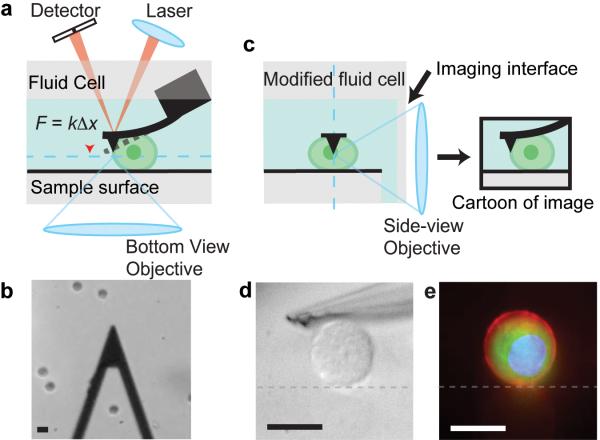
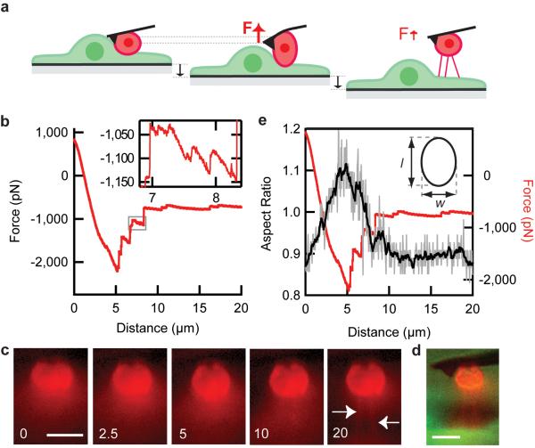
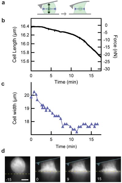
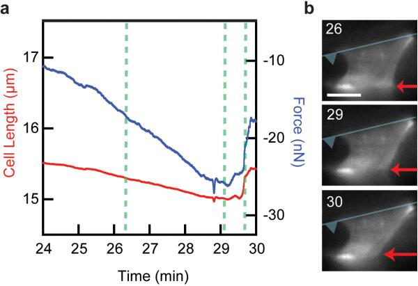
The mechanical rigidity of cells and adhesion forces between cells are important in various biological processes, including cell differentiation, proliferation and tissue organization. Atomic force microscopy has emerged as a powerful tool to quantify the mechanical properties of individual cells and adhesion forces between cells. Here we demonstrate an instrument that combines atomic force microscopy with a side-view fluorescent imaging path that enables direct imaging of cellular deformation and cytoskeletal rearrangements along the axis of loading. With this instrument, we directly observed cell shape under mechanical load, correlated changes in shape with force-induced ruptures and imaged formation of membrane tethers during cell-cell adhesion measurements. Additionally, we observed cytoskeletal reorganization and stress-fiber formation while measuring the contractile force of an individual cell. This instrument can be a useful tool for understanding the role of mechanics in biological processes.
Figures
References
-
- Engler AJ, Sen S, Sweeney HL, Discher DE. Matrix elasticity directs stem cell lineage specification. Cell. 2006;126:677–689. - PubMed
-
- Paszek MJ, et al. Tensional homeostasis and the malignant phenotype. Cancer Cell. 2005;8:241–254. - PubMed
-
- Krieg M, et al. Tensile forces govern germ-layer organization in zebrafish. Nature Cell Biology. 2008;10:429–U122. - PubMed
Publication types
MeSH terms
Substances
Grants and funding
LinkOut - more resources
Full Text Sources
Other Literature Sources
