. 2009 May;15(5):559-65.
doi: 10.1038/nm.1944. Epub 2009 Apr 12.

Copy number analysis indicates monoclonal origin of lethal metastatic prostate cancer

Copy number analysis indicates monoclonal origin of lethal metastatic prostate cancer

Wennuan Liu et al. Nat Med. 2009 May.

Erratum in

  • Nat Med. 2009 Jul;15(7):819

Abstract

Many studies have shown that primary prostate cancers are multifocal and are composed of multiple genetically distinct cancer cell clones. Whether or not multiclonal primary prostate cancers typically give rise to multiclonal or monoclonal prostate cancer metastases is largely unknown, although studies at single chromosomal loci are consistent with the latter case. Here we show through a high-resolution genome-wide single nucleotide polymorphism and copy number survey that most, if not all, metastatic prostate cancers have monoclonal origins and maintain a unique signature copy number pattern of the parent cancer cell while also accumulating a variable number of separate subclonally sustained changes. We find no relationship between anatomic site of metastasis and genomic copy number change pattern. Taken together with past animal and cytogenetic studies of metastasis and recent single-locus genetic data in prostate and other metastatic cancers, these data indicate that despite common genomic heterogeneity in primary cancers, most metastatic cancers arise from a single precursor cancer cell. This study establishes that genomic archeology of multiple anatomically separate metastatic cancers in individuals can be used to define the salient genomic features of a parent cancer clone of proven lethal metastatic phenotype.


Figures

Figure 1
Figure 1
a. Metastatic prostate cancer study subjects. Anatomic sample type indicators for 85 cancerous DNA samples studied by CGH and 58 cancerous samples studied by Affy6 are superimposed on posterior bone scan views for each subject. Legend indicates color and number coding of anatomic origin categories. Subjects from whom two or more anatomically distinct prostate cancer samples were studied are denoted with colored symbols upon which the subject’s number is superimposed. b. Unsupervised hierarchical clustering of cCGH data. Unsupervised hierarchical clustering dendrogram based on SAM-reduced 218 locus metastatic prostate cancer cCGH dataset for 80 samples from 24 subjects in which more than one anatomically separate cancerous DNA sample was available. All samples from an individual subject are color/shape coded using symbols shown (Fig. 1a). c. Discriminatory Component Analysis of cCGH data. Weighted Fisher criterion based discriminatory component analysis (wFC-DCA) of cCGH data from 80 metastatic prostate cancer samples from 24 subjects projected in 3-D Euclidean space. Samples are identified using color/shape symbols indicated (Fig 1a). d. Discriminatory Component Analysis of Affy6 data. Weighted Fisher criterion based discriminatory component analysis (wFC-DCA) of Affy6 data from 58 metastatic prostate cancer samples from 14 subjects projected in 3-D Euclidean space. Samples are identified using color/shape symbols indicated (Fig 1a). e. Unsupervised Hierarchical Clustering of Affy6 copy number data from 58 anatomically separate metastatic prostate cancer sites in 14 subjects. All samples from each of 14 subjects cluster together. Of 3,029,978 total genome segments analyzed in 58 samples studied (52241 per sample), 52.4% show no change in copy number vs subject-specific normal control baseline, 25.4% show gain, and 22.2% show loss.
Figure 2a
Figure 2a. Representative sample of allele-specific copy number data from subject 17 (chromosomes 6 and 13)
2b: Changes present in all samples are termed omniclonal (circled in green). 2c: Subclonal changes are present in just one (dark blue), a pair (brown) or three of the samples (light blue) studied. On chromosomes where whole chromosome arm gain or loss appears to have occurred before or after complex intrachromosomal gains and losses took place are difficult to interpret with existing technology and are labeled indeterminate (pink). 2d: Summary of all changes detected. 2e: Depiction of detected changes at each metastatic site studied, superimposed on posterior bone scan view with tan circle representing prostate, black circle representing cancer capable of metastasis. The parent cancer cell arises in the prostate and contains a set of clonally maintained copy number changes depicted in green at the base of each triangle containing labeled changes identified in each metastatic cancer sample. The parent cancer cell divides, maintaining the original set of changes but also giving rise to some new changes that are also maintained subclonally in cells that leave the prostate to populate various metastatic sites which were analyzed in this study. Subclonal diversification likely occurs in both the prostate and sites after initial metastasis, the simplest possible case (subclonal diversification in the prostate) is depicted for discussion purposes only, subclonal diversification likely occurs both in the prostate and at metastatic sites.
Figure 3a
Figure 3a. Representative sample of allele-specific copy number data from subject 34 (chromosomes 5 and 8)
3b: Changes present in all samples are termed omniclonal (circled in green). 3c: Subclonal changes are present in just one (dark blue), a pair (brown) or three of the samples (light blue) studied. Indeterminate changes are circled in pink. 3d: Summary of all changes detected. 3e: Depiction of detected changes at each metastatic site studied, superimposed on posterior bone scan view with tan circle representing prostate, black circle representing cancer capable of metastasis.
Figure 4
Figure 4. Potential patterns of metastatic prostate spread
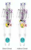
Our results show that most if not all metastatic prostate cancers have clonal origins. Using subject A17 as representative of all subjects, and considering recent data suggesting that in some men prostate cancer cells may lie dormant in the bone marrow for many years, , spread of cancer cells with common clonal origins occurs in a “Direct Clonal” or “Indirect Clonal” pattern shown. The large tan/pink circle represents the prostate, and the black circle represents prostate cancers capable of lethal spread. Green circles represent local prostate cancers incapable of spread, and Yellow circles represent nonlethal spreading cancer as suggested by data from Ellis et al. We found no significant difference in copy number patterns in prostate cancer foci isolated from the prostate at autopsy and metastases from various sites in the 5 subjects where prostate cancer foci were isolated from the prostate at autopsy. “Direct clonal” lethal metastasis provides the simplest explanation of these findings, since “Indirect” metastasis would require that the metastatic prostate cancer metastasize back to the prostate as illustrated by the dashed arrows.

Comment in

Similar articles

Cited by

References

    1. Miller GJ, Cygan JM. Morphology of prostate cancer: the effects of multifocality on histological grade, tumor volume and capsule penetration. Journal of Urology. 1994;152:1709–1713. [see comments] - PubMed
    1. Ruijter ET, van de Kaa CA, Schalken JA, Debruyne FM, Ruiter DJ. Histological grade heterogeneity in multifocal prostate cancer. Biological and clinical implications. J Pathol. 1996;180:295–299. - PubMed
    1. Aihara M, Wheeler TM, Ohori M, Scardino PT. Heterogeneity of prostate cancer in radical prostatectomy specimens. Urology. 1994;43:60–66. - PubMed
    1. Cheng L, et al. Evidence of independent origin of multiple tumors from patients with prostate cancer. Journal of the National Cancer Institute. 1998;90:233–237. - PubMed
    1. Macintosh CA, Stower M, Reid N, Maitland NJ. Precise microdissection of human prostate cancers reveals genotypic heterogeneity. Cancer Research. 1998;58:23–28. - PubMed

Publication types

MeSH terms