. 2009 Sep;58(9):1471-80.
doi: 10.1007/s00262-009-0705-8. Epub 2009 Apr 12.

The new face of nucleolin in human melanoma

Affiliations

The new face of nucleolin in human melanoma

Dorota Hoja-Łukowicz et al. Cancer Immunol Immunother. 2009 Sep.

Abstract

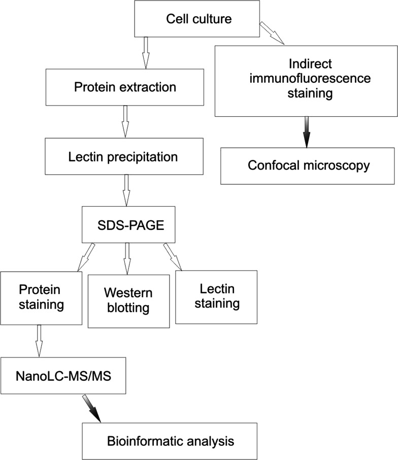
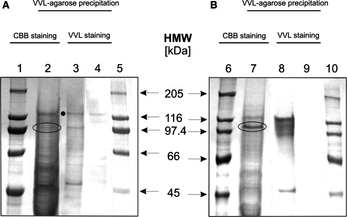
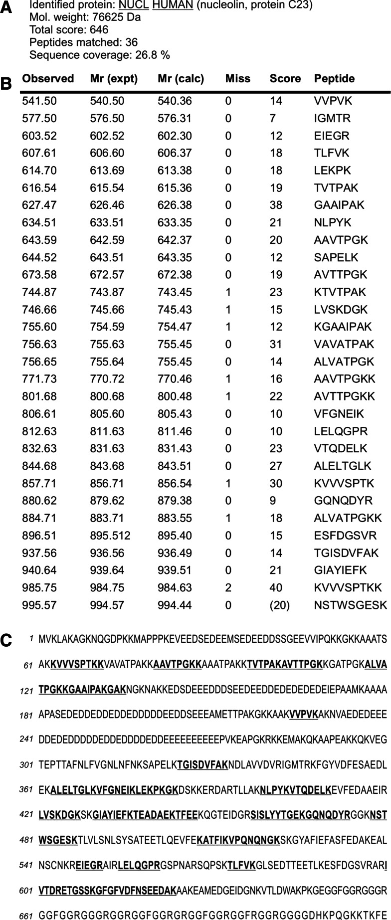
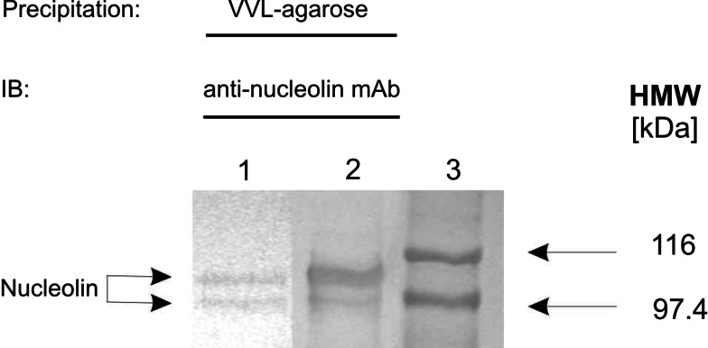
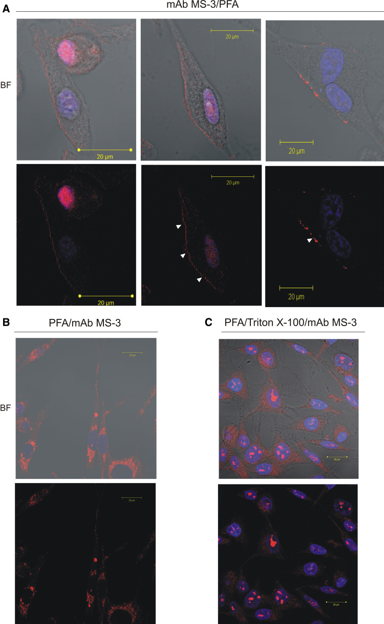
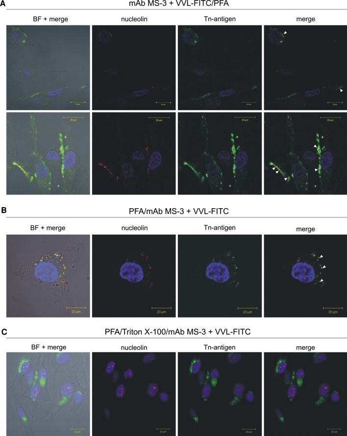
Nucleolin is multifunctional protein mainly present in nucleoli but also detected in cytoplasm and plasma membranes. Extranuclear nucleolin differs from the nuclear form by its glycosylation. Studies on expression of nucleolin in breast cancer suggest a possible association to the metastatic cascade. In the present study, Vicia villosa lectin (VVL) precipitation followed by subsequent polyacrylamide gel electrophoresis and mass spectrometry analysis demonstrates nucleolin as a VVL-positive glycoprotein expressed in melanoma. The presence of VVL-positive nucleolin in the melanoma cell membrane and cytoplasm was confirmed by confocal microscopy. Using bioinformatic peptide prediction programs, nucleolin was shown to contain multiple possible MHC class-I binding peptides in its sequence which makes nucleolin an interesting melanoma marker and target for immunodiagnostic and possibly therapeutic purposes.


Figures

Fig. 1
Fig. 1
Overview of the technique for analysis of Tn antigen-bearing glycoproteins
Fig. 2
Fig. 2
Lectin precipitation of Tn antigen-bearing glycoproteins in primary and metastatic melanoma. Extracts of WM793 (a) and Ma-Mel-27 (b) cellular proteins were precipitated using VVL-agarose, separated by SDS-PAGE and stained with CBB (lanes 2 and 7) or electrotransferred on PVDF membrane and probed with VVL (lanes 3 and 8) or VVL pre-blocked with GalNAc (lanes 4 and 9). In parallel, protein standards were resolved (lanes 1, 5, 6 and 10). Bands of interest (103 kDa for WM793 cell extract and 105 kDa for Ma-Mel-27 cell extract) were excised from the gel and subjected to mass spectrometry analysis (the encircled bands in lines 2 and 7). Note. One non-specific band was observed and marked with a black dot
Fig. 3
Fig. 3
Results of the mass spectrometry analysis of in-gel tryptic digest of 105 kDa protein band isolated from total lysate of Ma-Mel-27 cells using VVL-agarose. The encircled band shown in Fig. 2 lane 7 was excised from polyacrylamide gel and digested with trypsin, and the tryptic peptides were sequenced. The acquired spectra were analysed using Bruker Data Analysis software and interpreted using Mascot search engine against Swiss-Prot/TrEMBL sequence database. Peptides determined by sequencing were found to correspond to a high degree of certainty to human nucleolin (a). The table columns contain: Observed, experimental m/z value; Mr(expt), experimental m/z transformed to a relative molecular mass; Mr(calc), relative molecular mass calculated from the matched peptide sequence; Miss, number of missed cleavage sites; Score, ions score; Peptide, sequence of the matched peptide in 1-letter code (b). Localization of the identified peptides within the nucleolin sequence. The primary structure of human nucleolin is shown in the single-letter amino acid code sequence. Matched tryptic peptides are in bold and underlined (c). Note. Matched peptides equally cover the nucleolin sequence
Fig. 4
Fig. 4
Confirmation of mass spectrometry results. Extracts of WM793 (lane 1) and Ma-Mel-27 (lane 2) cellular proteins were precipitated using VVL-agarose, separated by SDS-PAGE and blotted on PVDF membrane. In parallel, protein standards were resolved (lane 3). Immunodetection of nucleolin was performed using anti-human nucleolin mAb, clone 3G4B2
Fig. 5
Fig. 5
Detection of membrane-associated and intracellular nucleolin in melanoma cells by confocal immunofluorescence laser microscopy. For the detection of the cell-surface-clustered nucleolin (a), Ma-Mel-27 cells cultured in fresh medium for 5 h were further incubated for 1 h at 37°C in the presence of mAb MS-3 diluted 1:25 in 10% NGS, 2% BSA/PBS containing 25 mM NaN3 before PFA fixation. For staining of semi-permeabilised (b) or permeabilised cells (c) Ma-Mel-27 cells (after 5 h of culture in fresh medium) were fixed with 3% PFA or 2% PFA/Triton before incubation with mAb MS-3 in 1% BSA/PBS (overnight, RT) diluted 1:100, respectively. The bound anti-nucleolin antibody was revealed by Cy3-labeled goat anti-mouse antibodies (red). Nuclei were counterstained with DAPI (blue). BF; bright field. Note. In PFA/Triton-fixed cells, nucleolin was detected primarily as fine dot-like structures in the nuclei and also as spots in the cytoplasm (c). In cells preincubated with anti-nucleolin mAb (living unpermabilised cells) red patches indicate cell surface nucleolin (arrows in a) (color figure online)
Fig. 6
Fig. 6
Cell surface and cytoplasmic nucleolin are carrying Tn antigen. Living unpermeabilised melanoma Ma-Mel-27 cells (after 5 h of culture in fresh medium) were simultaneously incubated with anti-nucleolin mAb MS-3 (dilution 1:25) and with FITC-labeled VVL (green, dilution 1:100) in 10% NGS, 2% BSA/PBS containing 25 mM NaN3 before PFA fixation (a). For staining of semi-permeabilised (b) or permeabilised cells (c) Ma-Mel-27 cells (after 5 h of culture in fresh medium) were fixed with 3% PFA or 2% PFA/Triton before simultaneous incubation with mAb MS-3 (dilution 1:100) and with FITC-labeled VVL (green, dilution 1:100) in 1% BSA/PBS (overnight, RT), respectively. The bound anti-nucleolin antibody was revealed by Cy3-labeled goat anti-mouse antibodies (red). Nuclei were counterstained with DAPI (blue). Arrows indicate colocalization (yellow) of VVL with nucleolin present on the cell surface. BF; bright field (color figure online)

Similar articles

Cited by

References

    1. Aldi S, Della Giovampaola C, Focarelli R, Armini A, Ziche M, Finetti F, Rosati F. A fucose-containing O-glycoepitope on bovine and human nucleolin. Glycobiology. 2009;19:337–343. doi: 10.1093/glycob/cwn126. - DOI - PubMed
    1. Bubenik J. MHC class I down-regulation, tumour escape from immune surveillance and design of therapeutic strategies. Folia Biol (Praha) 2005;51:1–2. - PubMed
    1. Carpentier M, Morelle W, Coddeville B, Pons A, Masson M, Mazurie J, Legrand D. Nucleolin undergoes partial N- and O-glycosylations in the extracellular cell compartment. Biochemistry. 2005;44:5804–5815. doi: 10.1021/bi047831s. - DOI - PubMed
    1. Chen C, Chiang S, Yeh N. Increased stability of nucleolin in proliferating cells by inhibition of its self-cleaving activity. J Biol Chem. 1991;266:7754–7758. - PubMed
    1. Christian S, Pilch J, Akerman ME, Porkka K, Laakkonen P, Ruoslahti E. Nucleolin expressed at the cell surface is a marker of endothelial cells in angiogenic blood vessels. J Cell Biol. 2003;163:871–878. doi: 10.1083/jcb.200304132. - DOI - PMC - PubMed

Publication types

MeSH terms