Parcellation of human temporal polar cortex: a combined analysis of multiple cytoarchitectonic, chemoarchitectonic, and pathological markers
- PMID: 19363802
- PMCID: PMC3665344
- DOI: 10.1002/cne.22053
Parcellation of human temporal polar cortex: a combined analysis of multiple cytoarchitectonic, chemoarchitectonic, and pathological markers
Abstract
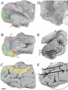
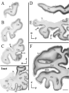
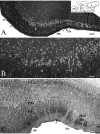
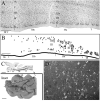
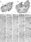
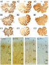
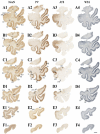
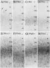
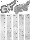
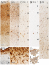
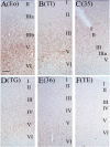
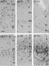
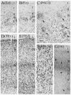
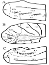
Although the human temporal polar cortex (TPC), anterior to the limen insulae, is heavily involved in high-order brain functions and many neurological diseases, few studies on the parcellation and extent of the human TPC are available that have used modern neuroanatomical techniques. The present study investigated the TPC with combined analysis of several different cellular, neurochemical, and pathological markers and found that this area is not homogenous, as at least six different areas extend into the TPC, with another area being unique to the polar region. Specifically, perirhinal area 35 extends into the posterior TPC, whereas areas 36 and TE extend more anteriorly. Dorsolaterally, an area located anterior to the typical area TA or parabelt auditory cortex is distinguishable from area TA and is defined as area TAr (rostral). The polysensory cortical area located primarily in the dorsal bank of the superior temporal sulcus, separate from area TA, extends for some distance into the TPC and is defined as the TAp (polysensory). Anterior to the limen insulae and the temporal pyriform cortex, a cortical area, characterized by its olfactory fibers in layer Ia and lack of layer IV, was defined as the temporal insular cortex and named as area TI after Beck (J. Psychol. Neurol. 1934;41:129-264). Finally, a dysgranular TPC region that capped the tip with some extension into the dorsal aspect of the TPC is defined as temporopolar area TG. Therefore, the human TPC actually includes areas TAr and TI, anterior parts of areas 35, 36, TE, and TAp, and the unique temporopolar area TG.
Figures

Similar articles
-
Perirhinal and parahippocampal cortices of the macaque monkey: cortical afferents.J Comp Neurol. 1994 Dec 22;350(4):497-533. doi: 10.1002/cne.903500402. J Comp Neurol. 1994. PMID: 7890828
-
Comparative neuroanatomical parcellation of the human and nonhuman primate temporal pole.J Comp Neurol. 2013 Dec 15;521(18):4163-76. doi: 10.1002/cne.23431. J Comp Neurol. 2013. PMID: 23881792 Review.
-
The abnormally phosphorylated tau lesion of early Alzheimer's disease.Neurochem Res. 2009 Jan;34(1):118-23. doi: 10.1007/s11064-008-9701-1. Epub 2008 Apr 25. Neurochem Res. 2009. PMID: 18437565
-
Cortical afferents to behaviorally defined regions of the inferior temporal and parahippocampal gyri as demonstrated by WGA-HRP.J Comp Neurol. 1992 Jul 8;321(2):177-92. doi: 10.1002/cne.903210202. J Comp Neurol. 1992. PMID: 1380012
-
Cortical connections of the inferior arcuate sulcus cortex in the macaque brain.Brain Res. 1992 Feb 21;573(1):8-26. doi: 10.1016/0006-8993(92)90109-m. Brain Res. 1992. PMID: 1374284
Cited by
-
Differences in Early Stages of Tactile ERP Temporal Sequence (P100) in Cortical Organization during Passive Tactile Stimulation in Children with Blindness and Controls.PLoS One. 2015 Jul 30;10(7):e0124527. doi: 10.1371/journal.pone.0124527. eCollection 2015. PLoS One. 2015. PMID: 26225827 Free PMC article.
-
Lesion symptom mapping of manipulable object naming in nonfluent aphasia: can a brain be both embodied and disembodied?Cogn Neuropsychol. 2014;31(4):287-312. doi: 10.1080/02643294.2014.914022. Cogn Neuropsychol. 2014. PMID: 24839997 Free PMC article.
-
Cellular resolution anatomical and molecular atlases for prenatal human brains.J Comp Neurol. 2022 Jan;530(1):6-503. doi: 10.1002/cne.25243. J Comp Neurol. 2022. PMID: 34525221 Free PMC article.
-
Lower brain glucose metabolism in normal ageing is predominantly frontal and temporal: A systematic review and pooled effect size and activation likelihood estimates meta-analyses.Hum Brain Mapp. 2023 Feb 15;44(3):1251-1277. doi: 10.1002/hbm.26119. Epub 2022 Oct 21. Hum Brain Mapp. 2023. PMID: 36269148 Free PMC article.
-
Predicting the location of human perirhinal cortex, Brodmann's area 35, from MRI.Neuroimage. 2013 Jan 1;64:32-42. doi: 10.1016/j.neuroimage.2012.08.071. Epub 2012 Aug 30. Neuroimage. 2013. PMID: 22960087 Free PMC article.
References
-
- Afifi AK, Bergman PA. Functional neuroanatomy: test and atlas. McGraw-Hill Companies; New York: 1998.
-
- Ang LC, Munoz DG, Shul D, George DH. SMI-32 immunoreactivity in human striate cortex during postnatal development. Dev Brain Res. 1991;61:103–109. - PubMed
-
- Arnold SE, Hyman BT, Flory J, Damasio AR, Van Hoesen GW. The topographical and neuroanatomical distribution of neurofibrillary tangles and neuritic plaques in the cerebral cortex of patients with Alzheimer’s disease. Cereb Cortex. 1991;1:103–116. - PubMed
-
- Arnold SE, Hyman BT, Van Hoesen GW. Neuropathologic changes of the temporal pole in Alzheimer’s disease and Pick’s disease. Arch Neurol. 1994;51:145–50. - PubMed
Publication types
MeSH terms
Substances
Grants and funding
LinkOut - more resources
Full Text Sources
Miscellaneous

