

A new dynamic 3D virtual methodology for teaching the mechanics of atrial septation as seen in the human heart
- PMID: 19363807
- PMCID: PMC2702359
- DOI: 10.1002/ase.74
A new dynamic 3D virtual methodology for teaching the mechanics of atrial septation as seen in the human heart
Abstract
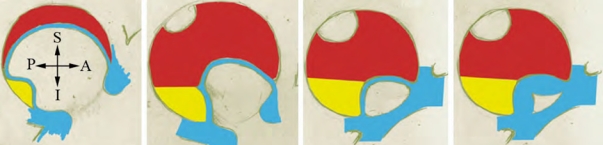
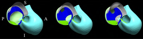
Learning embryology remains difficult, since it requires understanding of many complex phenomena. The temporal evolution of developmental events has classically been illustrated using cartoons, which create difficulty in linking spatial and temporal aspects, such correlation being the keystone of descriptive embryology. We synthesized the bibliographic data from recent studies of atrial septal development. On the basis of this synthesis, consensus on the stages of atrial septation as seen in the human heart has been reached by a group of experts in cardiac embryology and pediatric cardiology. This has permitted the preparation of three-dimensional (3D) computer graphic objects for the anatomical components involved in the different stages of normal human atrial septation. We have provided a virtual guide to the process of normal atrial septation, the animation providing an appreciation of the temporal and morphologic events necessary to separate the systemic and pulmonary venous returns. We have shown that our animations of normal human atrial septation increase significantly the teaching of the complex developmental processes involved, and provide a new dynamic for the process of learning.
Figures


Similar articles
-
Web-based interactive 3D visualization as a tool for improved anatomy learning.Anat Sci Educ. 2009 Mar-Apr;2(2):61-8. doi: 10.1002/ase.76. Anat Sci Educ. 2009. PMID: 19363804
-
Impact of cardiovascular embryology animations on short-term learning.Adv Physiol Educ. 2019 Mar 1;43(1):55-65. doi: 10.1152/advan.00121.2018. Adv Physiol Educ. 2019. PMID: 30615476
-
Virtual cerebral ventricular system: an MR-based three-dimensional computer model.Anat Sci Educ. 2011 Nov-Dec;4(6):340-7. doi: 10.1002/ase.256. Epub 2011 Oct 4. Anat Sci Educ. 2011. PMID: 21976457
-
Demonstration of living anatomy clarifies the morphology of interatrial communications.Heart. 2018 Dec;104(24):2003-2009. doi: 10.1136/heartjnl-2018-313758. Epub 2018 Sep 4. Heart. 2018. PMID: 30181201 Review.
-
Building virtual models by postprocessing radiology images: A guide for anatomy faculty.Anat Sci Educ. 2010 Sep-Oct;3(5):261-6. doi: 10.1002/ase.175. Anat Sci Educ. 2010. PMID: 20827725 Review.
Cited by
-
Learning Cardiac Embryology-Which Resources Do Students Use, and Why?Med Sci Educ. 2019 Sep 13;29(4):1051-1060. doi: 10.1007/s40670-019-00803-4. eCollection 2019 Dec. Med Sci Educ. 2019. PMID: 34457583 Free PMC article.
-
Cor triatriatum sinister versus bowed septum primum in an infant with total anomalous pulmonary venous connection: a difficult imaging distinction.Pediatr Radiol. 2012 Oct;42(10):1254-8. doi: 10.1007/s00247-012-2382-6. Epub 2012 Apr 28. Pediatr Radiol. 2012. PMID: 22544301
-
Exploring Visualisation for Embryology Education: A Twenty-First-Century Perspective.Adv Exp Med Biol. 2022;1356:173-193. doi: 10.1007/978-3-030-87779-8_8. Adv Exp Med Biol. 2022. PMID: 35146622
-
Three dimensional modeling of atrioventricular valves provides predictive guides for optimal choice of prosthesis.Sci Rep. 2022 May 6;12(1):7432. doi: 10.1038/s41598-022-10515-2. Sci Rep. 2022. PMID: 35523789 Free PMC article.
-
The influence of learning style in understanding analogies and 2D animations in embryology course.Anat Cell Biol. 2018 Dec;51(4):260-265. doi: 10.5115/acb.2018.51.4.260. Epub 2018 Dec 29. Anat Cell Biol. 2018. PMID: 30637160 Free PMC article.
References
-
- Achtemeier SD, Morris LV, Finnegan CL. Considerations for developing evaluations of online courses. Journal of Asynchronous Learning Networks. 2003;7 URL: http://www.aln.org/publications/jaln/v7nl/v7nl_achtemeier.asp.
-
- Anderson RH, Brown NA, Moorman AF. Development and structures of the venous pole of the heart. Dev Dyn. 2006;235:2–9. - PubMed
-
- Berlage T. Augmented-reality communication for diagnostic tasks in cardiology. IEEE Trans Inf Technol Biomed. 1998;2:169–173. - PubMed
Publication types
MeSH terms
LinkOut - more resources
Full Text Sources
Medical
