. 2009 Jun 2;48(21):4476-87.
doi: 10.1021/bi8023509.

Crystal structure of Baeyer-Villiger monooxygenase MtmOIV, the key enzyme of the mithramycin biosynthetic pathway

Affiliations

Crystal structure of Baeyer-Villiger monooxygenase MtmOIV, the key enzyme of the mithramycin biosynthetic pathway

Miranda P Beam et al. Biochemistry. .

Abstract

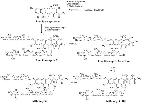
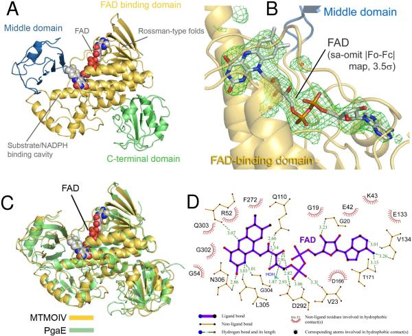
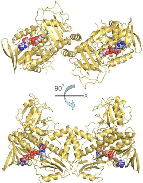
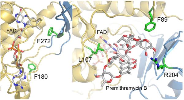
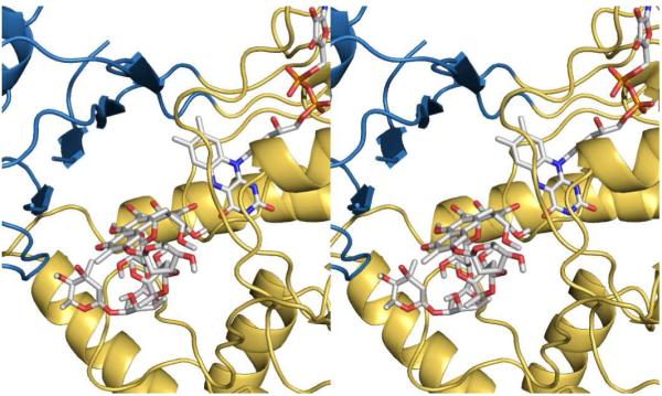
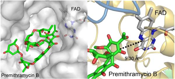
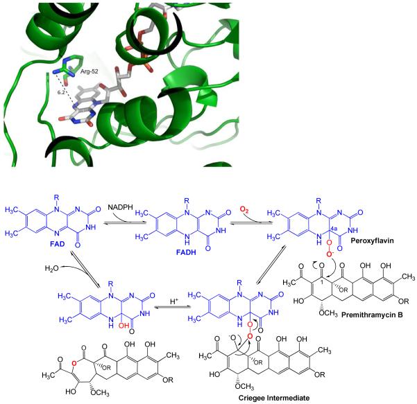
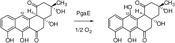
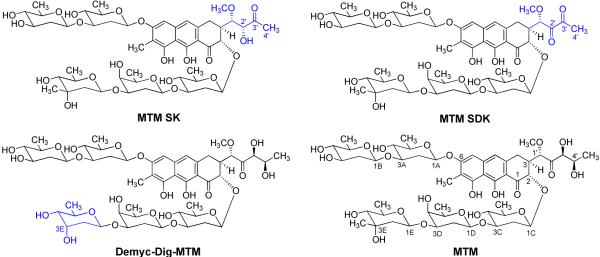
Baeyer-Villiger monooxygenases (BVMOs), mostly flavoproteins, were shown to be powerful biocatalysts for synthetic organic chemistry applications and were also suggested to play key roles for the biosyntheses of various natural products. Here we present the three-dimensional structure of MtmOIV, a 56 kDa homodimeric FAD- and NADPH-dependent monooxygenase, which catalyzes the key frame-modifying step of the mithramycin biosynthetic pathway and currently the only BVMO proven to react with its natural substrate via a Baeyer-Villiger reaction. MtmOIV's structure was determined by X-ray crystallography using molecular replacement to a resolution of 2.9 A. MtmOIV cleaves a C-C bond, essential for the conversion of the biologically inactive precursor, premithramycin B, into the active drug mithramycin. The MtmOIV structure combined with substrate docking calculations and site-directed mutagenesis experiments identifies several residues that participate in cofactor and substrate binding. Future experimentation aimed at broadening the substrate specificity of the enzyme could facilitate the generation of chemically diverse mithramycin analogues through combinatorial biosynthesis.


Figures

Figure 1
Figure 1
Biosynthetic pathway to mithramycin. The reaction catalyzed by BVMO MtmOIV yields premithramycin B-lactone, validating the BV mechanism, and is further converted to mithramycin DK (presumably spontaneously). The ketoreductase MtmW catalyzes the final step of mithramycin biosynthesis
Figure 2
Figure 2
A. Overall structure of MtmOIV. The FAD binding domain is shown in gold, the middle domain in blue, and the C-terminal domain in green. The FAD cofactor is shown as spheres. The Rossman-type fold and the substrate/co-factor binding cavity are indicated accordingly. B. View of FAD binding with FAD shown in stick figure and simulated annealing omit |Fo-Fc| electron density (3.5σ) for FAD in green mesh. The FAD binding domain is in gold and the middle domain is in blue. C. Superposition of the crystal structure of MtmOIV (gold) and PgaE (green). The FAD cofactor is shown as spheres. D. FAD binding with involved residues and distances. Figure was created using LIGPLOT (62).
Figure 3
Figure 3
Sequence alignment of MtmOIV, PgaE, and CabE. Green shading indicates residues that are completely conserved, yellow indicates partially conserved, and cyan indicates similar residues. Secondary α-helix structures are labeled as orange blocks above the residues, and β-sheets with purple arrows. Structural labels have the same numbering as the PgaE structure (44).
Figure 4
Figure 4
Biological subunit of MtmOIV as a dimer, which has ∼1200 Å2 of buried surface area (5.7% of total surface area for each monomer) which is mediated along the FAD binding domain and the middle domain.
Figure 5
Figure 5
A. Residues F180 (green), close to the adenine moiety of FAD, and F272 (green), close to the isoalloxazine moiety of FAD (gray stick with N atoms = blue, O-atoms = red and P atoms = orange), were selected for mutagenesis to support the FAD binding area in MtmOIV (gold/blue ribbon). B. Residues L107, R204 and F89 (all in green) were chosen to support their involvement in the substrate (premithramycin B, gray stick with O-atoms = red) binding. The isoalloxazine moiety of FAD (gray stick with N-atoms = blue, O-atoms = red, P-atoms = orange) and the enzyme show in gold/blue ribbon. The results (Table 2) show that residues F89 and R204 possibly constrict the premithramycin B binding site, while the L107A mutation reduces substrate binding, and also FAD binding.
Figure 6
Figure 6
Stereoimage of the premithramycin B (gray stick) binding pocket with FAD (gray stick), based on the found density from premithramycin B/MtmOIV co-crystallization and initial Autodock computational studies, prior to refinement (for the Autodock-refined results, see Figs. 5B and 7). MtmOIV is shown as gold (FAD binding domain) and blue (middle domain) ribbon.
Figure 7
Figure 7
A. Premithramycin B (green stick) binding pocket with FAD (gray stick). MtmOIV is shown in transparent gray surface representation. B. The dotted line represents the measured distance of 5.30 Å from the C4a carbon of the flavin ring of FAD to C1 of premithramycin B.
Figure 8
Figure 8
A. Left: View of the catalytically important arginine-52 (R52, green stick, N = blue, O = red) residue of MtmOIV above the flavin ring of FAD (gray stick, N = blue, O = red, P = orange). The measured distance (dotted line) between the N-atoms of the guanidine residue of R52 and the flavin ring surface is ∼ 6.2 Å in the shown conformation. The remainder of the enzyme (ribbon) is depicted in green. B. Below: Suggested Baeyer-Villiger reaction mechanism of the MtmOIV reaction, involving the peroxyflavin and Criegee intermediates. R = deoxysugar chains
Figure 9
Figure 9
The PgaE reaction. PgaE is an early acting hydroxylase in the biosynthesis of angucylinone gaudimycin C, catalyzing the 12-hydroxylation of UWM6 (left) to 12-hydroxy-UWM6 (right).
Figure 10
Figure 10
Chemical structures of mithramycin (MTM) analogues mithramycin SK (MTM SK)(40), mithramycin SDK (MtmSDK)(20) and 3D-demycarosyl-3D-digitoxosyl-mithramycin (Demyc-Dig-MTM)(41), all generated by combinatorial biosynthesis, which have increased efficacy compared with MTM. The structural differences compared with MTM are highlighted in blue.

Similar articles

Cited by

References

    1. Mihovilovic MD, Rudroff F, Grotzl B, Kapitan P, Snajdrova R, Rydz J, Mach R. Family clustering of Baeyer-Villiger monooxygenases based on protein sequence and stereopreference. Angew. Chem. Int. Ed. Engl. 2005;44:3609–3613. - PubMed
    1. van Berkel WJH, Kamerbeek NM, Fraaije MW. Flavoprotein monooxygenases, a diverse class of oxidative biocatalysts. J. Biotechnol. 2006;124:670–689. - PubMed
    1. Kamerbeek NM, Fraaije MW, Janssen DB. Identifying determinants of NADPH specificity in Baeyer-Villiger monooxygenases. Eur. J. Biochem. 2004;271:2107–2116. - PubMed
    1. Kamerbeek NM, Janssen DB, van Berkel WJH, Fraaije MW. Baeyer-Villiger Monooxygenases, and Emerging Family of Flavin-Dependent Biocatalysts. Adv. Synth. Catal. 2003;345:667–678.
    1. Willetts A. Structural studies and synthetic applications of Baeyer-Villiger monooxygenases. Trends Biotechnol. 1997;15:55–62. - PubMed

Publication types

Associated data