

doi: 10.1016/j.febslet.2009.04.007.
Epub 2009 Apr 11.
Endoplasmic reticulum stress is implicated in retinal inflammation and diabetic retinopathy
Affiliations
- PMID: 19364508
- PMCID: PMC2691649
- DOI: 10.1016/j.febslet.2009.04.007
Item in Clipboard
Endoplasmic reticulum stress is implicated in retinal inflammation and diabetic retinopathy
FEBS Lett.
.
Abstract
Diabetic retinopathy is a chronic low-grade inflammatory disease; however, the mechanisms remain elusive. In the present study, we demonstrated that endoplasmic reticulum (ER) stress was activated in the retina in animal models of diabetes and oxygen-induced retinopathy (OIR). Induction of ER stress by tunicamycin resulted in significantly increased expression of inflammatory molecules in the retina. Inhibition of ER stress by chemical chaperone 4-phenyl butyric acid ameliorated inflammation in cultured human retinal endothelial cells exposed to hypoxia, and in the retinas of diabetic and OIR mice. These findings indicate that ER stress is a potential mediator of retinal inflammation in diabetic retinopathy.
Figures

A). Western blot analysis of TNF-α and VEGF in the retinas of Akita mice. The membrane was reblotted with β-actin antibody as loading control. B). Retinal levels of TNF-α and VEGF were quantified by densitometry from 4 individual animals (mean ± SD, n = 4). C). mRNA level of GRP78 in the retina determined by real-time RT-PCR and normalized by 18S (mean ± SD, n = 7 in control group and n = 4 in Akita group). D). Representative images of GRP 78 expression in the retinas from 4 Akita mice and 5 littermate controls. Note more intensive signal of GRP 78 (brown color) in the inner retina of Akita mice (C-b) when compared to control (C-a). Magnification: 200x. ONL: outer nuclear layer; INL: inner nuclear layer; GCL: ganglion cell layer. E). Western blot analysis of phospho-IRE1α, phospho-eIF2α and ATF4 in the retinas of Akita mice. E). Retinal levels of phospho-IRE1α, phospho-eIF2α and ATF4 were quantified by densitometry (mean ± SD, n = 8). * p<0.05 and **p<0.01.

A). mRNA level of GRP78 in the retina of OIR mice determined by real-time RT-PCR and normalized by 18S (mean ± SD, n = 5). B). Protein level of GRP78 determined by Western blot analysis. Lower panel: densitometry results (mean ± SD, n = 8). C). Western blot analysis of phospho-eIF2α and ATF4 in the retina. D). Retinal levels of phospho-eIF2α and ATF4 were quantified by densitometry (mean ± SD, n=8). **p<0.01.
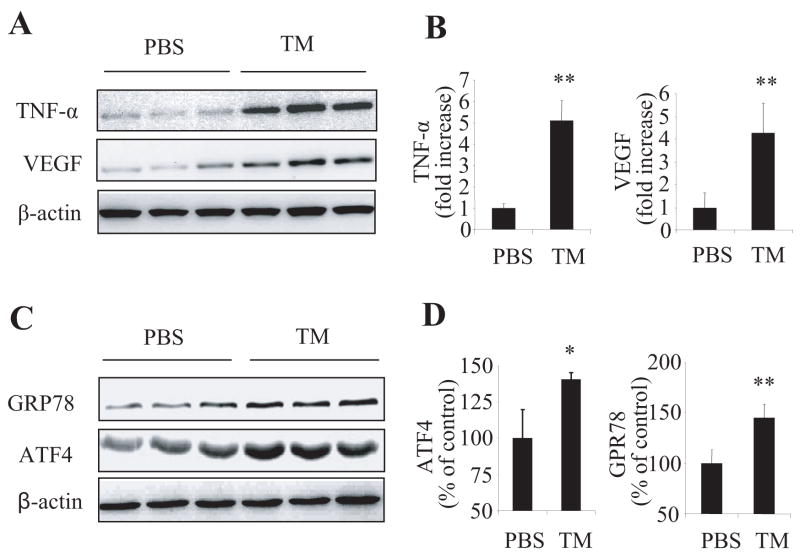
Adult C57BL/6J mice received periocular injection of tunicamycin in one eye and PBS as control in the contralateral eye. Twenty four hours after injection, retinas were dissected and expression of pro-inflammatory factors (TNF-α and VEGF) (A, B) and ER stress markers (GRP78 and ATF4) (C, D) were determined by Western blot analysis and quantified by densitometry (mean ± SD, n = 4). * p<0.05 and **p<0.01.

A). HREC were treated with hypoxia (2% O2) for 16h and GPR78 expression was determined by immunostaining. A-a: control; A-b: hypoxia. B–E). HREC were pre-treated with PBA at the doses as indicated for 8h and then exposed to hypoxia (2% O2) for 16h. B–C). Expression of phospho-IRE1α, phospho-eIF2α, and ATF4 was determined by Western blot analysis and quantified by densitometry (mean ± SD, n= 3) D–E). Expression of VEGF and TNF-α was measured by Western blot analysis and quantified by densitometry (mean ± SD, n = 3). The values statistically different from control were indicated as ** p<0.01; from hypoxia indicated as † p<0.05, ‡ p<0.01.

A). PBA (0.4 μmol/eye) was injected periocularly into one eye and same amount of vehicle was injected into the counterlateral eye as control in the Akita mice, twice a week, for 6 weeks. Expression of VEGF in the retina was measured by Western blot analysis and quantified by densitometry (mean ± SD, n = 4). B). OIR mice received intraperitoneal injection of PBA (40 mg/kg/body weight/day) or PBS from P7 to P14. Retinal VEGF expression was measured by Western blot analysis at P15 (mean ± SD, n = 5). * p<0.05 and ** p<0.01.
References
-
- Frank RN. Diabetic retinopathy New England. Journal of Medicine. 2004;350:48–58. - PubMed
-
- Zhang SX, Wang JJ, Gao G, Shao C, Mott R, Ma J-x. Pigment epithelium-derived factor (PEDF) is an endogenous anti-inflammatory factor. FASEB J. 2006;20:323–325. - PubMed
-
- Mitamura Y, Takeuchi S, Matsuda A, Tagawa Y, Mizue Y, Nishihira J. Monocyte chemotactic protein-1 in the vitreous of patients with proliferative diabetic retinopathy. Ophthalmologica. 2001;215:415–418. - PubMed
Publication types
MeSH terms
Substances
Grants and funding
LinkOut - more resources
Full Text Sources
Other Literature Sources
Medical
Molecular Biology Databases
