

Farnesol-induced apoptosis in Candida albicans
- PMID: 19364863
- PMCID: PMC2687256
- DOI: 10.1128/AAC.01551-08
Farnesol-induced apoptosis in Candida albicans
Abstract
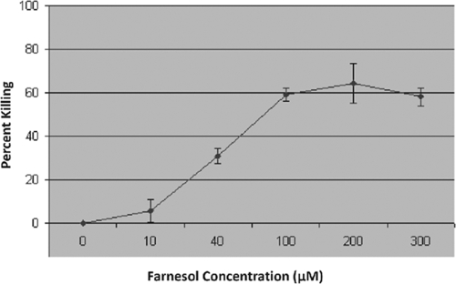
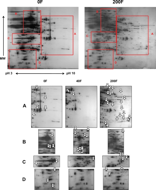
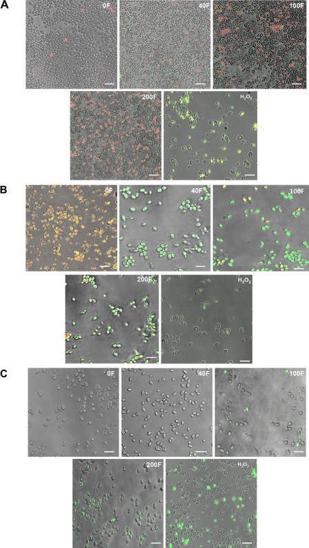
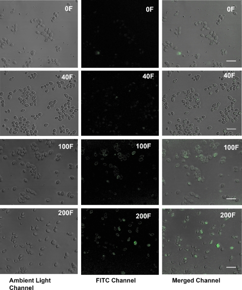
Farnesol, a precursor in the isoprenoid/sterol pathway, was recently identified as a quorum-sensing molecule produced by the fungal pathogen Candida albicans. Farnesol is involved in the inhibition of germination and biofilm formation by C. albicans and can be cytotoxic at certain concentrations. In addition, we have shown that farnesol can trigger apoptosis in mammalian cells via the classical apoptotic pathways. In order to elucidate the mechanism behind farnesol cytotoxicity in C. albicans, the response to farnesol was investigated, using proteomic analysis. Global protein expression profiles demonstrated significant changes in protein expression resulting from farnesol exposure. Among the downregulated proteins were those involved in metabolism, glycolysis, protein synthesis, and mitochondrial electron transport and the respiratory chain, whereas proteins involved in folding, protection against environmental and oxidative stress, actin cytoskeleton reorganization, and apoptosis were upregulated. Cellular changes that accompany apoptosis (regulated cell death) were further analyzed using fluorescent microscopy and gene expression analysis. The results indicated reactive oxygen species accumulation, mitochondrial degradation, and positive terminal deoxynucleotidyltransferase-mediated dUTP-biotin nick end labeling (TUNEL) in the farnesol-exposed cells concurrent with increased expression of antioxidant-encoding and drug response genes. More importantly, the results demonstrated farnesol-induced upregulation of the caspase gene MCA1 and the intracellular presence of activated caspases. In conclusion, this study demonstrated that farnesol promotes apoptosis in C. albicans through caspase activation, implying an important physiological role for farnesol in the fungal cell life cycle with important implications for adaptation and survival.
Figures
References
-
- Costa, V., and P. Moradas-Ferreira. 2001. Oxidative stress and signal transduction in Saccharomyces cerevisiae: insights into ageing, apoptosis and diseases. Mol. Aspects Med. 22:217-246. - PubMed
-
- Enari, M., H. Sakahira, H. Yokoyama, K. Okawa, A. Iwamatsu, and S. Nagata. 1998. A caspase-activated DNase that degrades DNA during apoptosis, and its inhibitor ICAD. Nature 391:43-50. - PubMed
Publication types
MeSH terms
Substances
Grants and funding
LinkOut - more resources
Full Text Sources
Molecular Biology Databases
